Fig. 2.1
A table-mounted slit-lamp biomicroscope offers excellent optics with multiple options for magnification and greater numbers of options for varying slit-beam width, height, and light color, and cameras may be added to the configuration to provide greater potential for stability and photographic documentation of lesions than camera options on handheld portable slit lamps
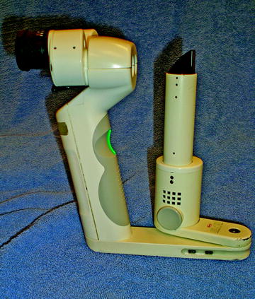
Fig. 2.2
The Kowa handheld slit lamp offers good optics and portability for the examination of eye in animals that are not sedated or that cannot be easily presented for examination by a table-mounted slit-lamp biomicroscope
Interpretation of the findings on slit-lamp biomicroscopy requires extensive knowledge of normal findings as well as background lesions that occur as incidental findings in the species and breed examined. These include, but are not limited to, embryonic remnants (e.g., persistent pupillary membranes, persistent or absorbing hyaloid arteries), corneal opacities (e.g., corneal scars, corneal degeneration/dystrophy in Sprague-Dawley rats, epithelial dystrophy in Dutch belted rabbits), cataracts, and lesions that may be associated with incidental trauma or inflammation (e.g., synechiae, traumatic cataracts, corneal scars).[1, 2, 10, 16–19] Examples of normal and abnormal slit-lamp findings are noted in Figs. 2.3, 2.4, 2.5, 2.6, 2.7, 2.8, and 2.9. Whenever possible, animals with such background lesions noted at pre-study examinations should not be placed on study. However, in some animals such as the Sprague-Dawley rat, the incidental lesions may be so pervasive that use of affected animals cannot be avoided and randomization will usually ensure the lesions are seen with equal representation across all groups. It is then important to ensure the test article administration does not result in worsening of the lesions. Testing in more than one species is the norm for topical ocular studies and provides additional confirmation of safety.
When slit-lamp examinations are used in systemic toxicology studies, it may be sufficient to simply record abnormal findings, but in topical ocular studies, it is customary to quantify findings and modifications of scoring systems such as the Hackett-McDonald scoring system (Table 2.1) [8, 11] and others such as outlined in the Standardization of Uveitis Nomenclature or SUN system [15]. Such scoring systems are designed to detect very subtle microscopic ocular changes, and they may be commonly employed in both preclinical and clinical studies. When evaluating the lens, the examination should include the notation of whether the lens is normal or abnormal and should be accompanied by a comment describing the extent and location of the lens abnormalities as determined by direct and indirect (i.e., retroillumination) illumination. Cataracts may be noted as mild (or incipient involving less than 10% of the lens), moderate (immature), or severe (involving the entire lens), and the location of the opacities may be described or defined by where they are localized in the lens (noted by the positions of the opacities in the slit beam as the light passes through the lens). Localization may thus be defined as anterior capsular, anterior subcapsular, anterior cortical, nuclear, posterior cortical, posterior subcapsular, posterior capsular, or equatorial with combinations of the preceding used if there is more than one opacity. In some instances, drawing of the lens with frontal and cross-sectional views may be used to further document the opacities. With such data recorded, it is thus possible to determine if a cataract is progressing during the study.
Table 2.1
Summary of a modified Hackett-McDonald ocular scoring system
Conjunctival congestion | |
Normal (0) = | Normal in appearance for the species. May appear blanched to reddish pink without perilimbal injection (except at 12 and 6 o’clock positions) with vessels of the palpebral and bulbar conjunctiva defined and easily observed |
1+ = | Mild. A flushed, reddish color predominantly confined to the palpebral conjunctiva with some perilimbal injection, primarily located but not confined to the upper and lower regions of the eye |
2+ = | Moderate. The palpebral conjunctiva appears bright red with accompanying perilimbal injection covering at least 75% of the circumference of the perilimbal region |
3+ = | Severe. Both the bulbar and palpebral conjunctiva exhibit a dark, beefy-red color with pronounced perilimbal injection. Petechiae on the conjunctiva may be present. The petechiae generally predominate along the nictitating membrane and/or the upper palpebral conjunctiva |
Conjunctival swelling | |
Normal (0) = | No swelling of the conjunctival tissue is observed |
1+ = | Minimal. Swelling above normal but without eversion of the eyelids. Swelling generally begins in the lower cul-de-sac near the inner canthus |
2+ = | Mild. Swelling with misalignment of the normal approximation of the lower and upper eyelids. In this stage, swelling is confined generally to the upper eyelid, with some swelling observed in the lower cul-de-sac |
3+ = | Moderate. Swelling is definite, with partial eversion of the upper and lower eyelids essentially equivalent. Eversion of the eyelids may be determined by looking at the animal head-on and observing the positioning of the eyelids |
4+ = | Severe. Eversion of the upper eyelid is pronounced with less pronounced eversion of the lower eyelid. At this level, it is difficult to retract the lids and observe the perilimbal region |
Conjunctival discharge | |
Normal (0) = | May include a small amount of clear, mucoid material that is normally found in the medial canthus of a substantial number of animal eyes |
1+ = | Mild. Discharge is above normal and present on the inner portion of the eye but not on the lids or hairs of the eyelids |
2+ = | Moderate. Discharge is abundant, easily observed, and has collected on the lids and around the hairs of the eyelids |
3+ = | Severe. Discharge has been flowing over the eyelids so as to substantially wet the hairs on the skin around the eye |
Iris congestion | |
Normal (0) = | Normal iris without any hyperemia of the iris vessels. Note: In rabbits, around the 12 to 1 o’clock position and the 6 to 7 o’clock position near the pupillary border, there may be a small area (approximately 1–3 mm in diameter) in which both the secondary and tertiary vessels may be slightly hyperemic. This is normal |
1+ = | Minimal. Minimal injection of secondary and tertiary vessels observed. Generally, it is uniform, but may be of greater intensity at the 12 to 1 o’clock or 6 o’clock position. If it is confined to this area, the tertiary vessels must be substantially hyperemic |
2+ = | Mild injection of tertiary vessels and minimal to moderate injection of the secondary vessels observed |
3+ = | Moderate injection of the secondary and tertiary vessels with slight swelling of the iris stroma (this gives the iris surface a slightly rugose appearance, which is usually most predominant near the 3 and 9 o’clock positions) |
4+ = | Severe. Marked injection of the secondary and tertiary vessels with definite swelling of the iris stroma. The iris appears rugose and may be accompanied by hemorrhage in the anterior chamber |
Pupillary light reflex | |
Normal (0) = | Normal pupillary light reflex |
1+ = | Sluggish pupillary light reflex |
2+ = | Maximally impaired pupillary reflex (pupil dilated and unresponsive) |
Aqueous flare | |
Normal = | Absence of visible light beam in the anterior chamber (no Tyndall effect) |
1+ = | Mild. The Tyndall effect is barely discernible. The intensity of the light beam in the anterior chamber is less than the intensity of the slit beam as it passes through the lens |
2+ = | Moderate. The Tyndall beam in the anterior chamber is easily discernible and is equal in intensity to the slit beam as it passes through the lens |
3+ = | Severe. The Tyndall beam in the anterior chamber is easily discernible; its intensity is greater than the intensity of the slit-lamp beam as it passes through the lens |
Corneal cloudiness severity | |
Normal = | Normal. Appears with the slit lamp as having a bright gray line on the epithelial surface and a bright gray line on the endothelial surface with a uniform marble-like gray appearance of the stroma |
1+ = | Minimal. Some loss of transparency. Only the epithelium and/or the anterior half of the stroma is involved as observed with an optical section of the slit lamp. With diffuse illumination, the underlying structures are clearly visible, although some cloudiness may be readily apparent |
2+ = | Mild. Some loss of transparency. The cloudiness extends past the anterior half of the stroma. The affected stroma has lost its marble-like appearance and is homogeneously white. With diffuse illumination, underlying structures are visible, although there may be some loss of detail |
3+ = | Moderate. Involvement of the entire thickness of the stroma. With optical section, the endothelial surface is still visible. However, with diffuse illumination, the underlying structures are just barely visible (to the extent that the observer is still able to grade flare and iritis, observe for pupillary response, and note lenticular changes) |
4+ = | Severe. Involvement of the entire thickness of the stroma. With optical section, the endothelium is not clearly visualized. With diffuse illumination, the underlying structures cannot be seen so that the evaluation of aqueous flare, iritis, pupillary response, and lenticular changes is not possible |
Ocular surface are involvement (applied to corneal clouding and staining of the cornea with fluorescein) | |
Normal (0) = | No area of cloudiness |
1+ = | Less than one fourth the corneal area |
2+ = | One fourth to less than one half the corneal area |
3+ = | One half to less than three fourths the corneal area |
4+ = | Three fourths or greater of the corneal area |
Pannus (corneal vascularization) | |
Normal (0) = | No pannus |
1+ = | Vascularization is present but vessels have not invaded the entire corneal circumference. Where localized vessel invasion has occurred, the vessels have not penetrated beyond 2 mm |
2+ = | Vessel invasion is greater than 2 mm in one or more areas or involves the entire corneal circumference |
Fluorescein staining intensity (area scored as above) | |
Normal (0) – | No staining. This may include a small number of faint focal or hazy areas of fluorescein staining which may be present in normal eyes |
1+ = | Slight fluorescein staining. With diffuse illumination, the underlying structures are easily visible. (The outline of the pupillary margin can be seen as if there were no fluorescein staining) |
2+ = | Mild fluorescein staining. With diffuse illumination, the underlying structures are visible, although there is some loss of detail |
3+ = | Marked fluorescein staining. With diffuse illumination, underlying structures are barely visible but not completely obscured |
4+ = | Severe fluorescein staining . With diffuse illumination, underlying structures cannot be observed |
Lens | |
Normal (N or 0) or abnormal (A or 1) – Describe the lenticular opacities and note position(s) in comments | |
In addition to evaluating the solid structures of the eye, the aqueous and anterior vitreous can also be evaluated as the slit beam passes through the anterior chamber and the anterior vitreous just posterior to the lens. Uveitis can result in an increase in protein and cells in the aqueous and vitreous in association with breakdown of the blood-ocular barrier. Increase in protein in the aqueous humor results in a Tyndall effect, referred to as aqueous flare, such that the light of the slit beam can be seen as it passes through the aqueous. As noted in Table 2.1, the flare may be scored from 0 to +3 where 0 equals the normal absence of a visible light beam as it passes through the aqueous, +1 equals a visible light beam that is less intense than the beam as it passes through the normal lens (this includes a barely visible beam which may be further described as trace flare), +2 equals a visible light beam equal in intensity to the beam as it passes through the normal lens, and +3 equals a visible light beam that is greater in intensity than the beam as it passes through the normal lens. As noted in the SUN system (Table 2.2), some systems adapt a slightly different mode for scoring aqueous flare to accommodate noting the presence or absence of fibrin in the aqueous. However, there can be varying amounts of fibrin formation, and some prefer to score fibrin formation in a class by itself (Table 2.3). Since the presence of fibrin in the anterior chamber generally indicates a greater degree of breakdown of the blood-aqueous barrier and hence greater inflammation, this latter practice allows better quantification if fibrin formation is encountered with any frequency in studies.
Table 2.2
SUNa grading scheme for aqueous flare
Grade | Description |
|---|---|
0 | None |
1+ | Faint |
2+ | Moderate (iris and lens details clear) |
3+ | Marked (iris and lens details hazy) |
4+ | Intense (fibrin or plasmoid aqueous) |
Table 2.3
Alternate scoring scheme for anterior chamber fibrin
Grade | Description (based upon degree of anterior chamber filled) |
|---|---|
0 | None |
1+ | Faint (<25% of anterior chamber) |
2+ | Mild (25–50% of anterior chamber) |
3+ | Moderate (51–75% of anterior chamber) |
4+ | Marked (>75% of anterior chamber) |
The presence of cells in the aqueous and anterior vitreous can be observed with slit-lamp biomicroscopy, and the SUN system developed a reasonable method for scoring the quantity of cells observed in the aqueous in a 1 X 1 mm beam (Table 2.4). The observer should concentrate on counting the cells at the same point (usually in the central anterior chamber) without moving the beam since cells are heavier than the surrounding aqueous and will settle inferiorly. In addition to noting the number of cells in the aqueous, it is important to note the types of cells as they may be pigmented arising from the uvea or retinal pigment epithelium (due to injury to or shedding from those structures), nonpigmented (white blood cells arising from an inflammatory response), or red blood cells (arising from damaged vasculature) as may occur with surgical or other invasive procedures (intraocular injections, aqueous or vitresous aspirations, lens extraction, placement of intraocular implants, subretinal injections, etc.). When nonpigmented cells are present in such numbers that frank hypopyon (pus in the anterior chamber) occurs, that should be noted as a comment in addition to the scoring [9, 15]. Likewise, when frank hemorrhage is noted (hyphema with or without clots), that too is worthy of separate comment and clarification.


Table 2.4
SUNa grading scheme for cells
< div class='tao-gold-member'>
Only gold members can continue reading. Log In or Register to continue
Stay updated, free articles. Join our Telegram channel
Full access? Get Clinical Tree
