Fig. 1.
Demonstrating the type of digital photo of the brain required for SAH grading. Basilar artery (BA), anterior cerebral artery (ACA), internal carotid artery (ICA), proximal posterior cerebral artery (pPCA), posterior communicating artery (PcomA), middle cerebral artery (MCA), distal posterior cerebral artery (dPCA), superior cerebellar artery (SCA). Photo adopted with modification from Sugawara et al. (11).
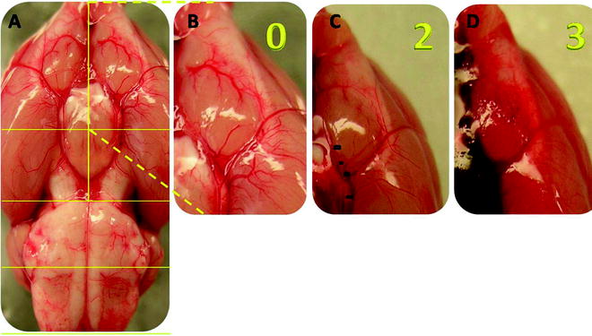
Fig. 2.
Example of the grading system using examples of the left frontal basilar cistern as an example. (a) Shows the brain from an SHAM animal. (b) Close up view of the left frontal basilar cistern shows clearly visible vessels without subarachnoid blood. The grade for this section is 0. (c) The left frontal basilar cistern from an SAH animal. This time the cistern has a significant blood clot but it does not obscure the ICA and ACA arteries. The grade for this cistern is 2. (d) Left frontal basilar cistern of another SAH animal. This time there is severe SAH, obliterating the view of all vessels. The grade here is 3. Photo adopted with modification from Sugawara et al. (11).
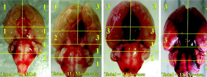
Fig. 3.
Here are four examples of various SAH grades ranging from mild to severe created using the endovascular perforation model in rats. The conditions for each animal were under identical and under controlled conditions. These photos represent the variability inherent in the endovascular perforation model.
3.2 Grading SAH Severity
Grading should be done by a blinded observer.
1.
We identified and used the arteries within the basal cistern for this grading system: basilar artery (BA) and the circle of Willis composed of the anterior cerebral artery (ACA), the internal carotid artery (ICA), the proximal posterior cerebral artery (pPCA) from ICA to posterior communicating artery (PcomA), and the PcomA. Middle cerebral artery (MCA), distal posterior cerebral artery (dPCA), and the superior cerebellar artery (SCA) were not included in the grading system. Only the arteries of the basal cisterns were included in the grading system to maintain reproducibility and consistency (Fig. 1).
3.
4.
The animals received a total score ranging from 0 to18 after adding the scores from all six segments (Fig. 3).
(a)
Based on the final score the animals are divided into three groups:
(b)
Grade 0–7: mild SAH
(c)
Grade 8–12: moderate SAH
(d)
Grade 13–18: severe SAH
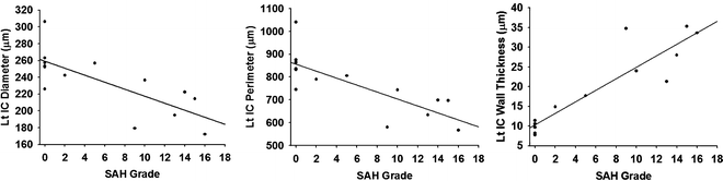
4 Outcome Measures that Have Proven to Correlate with this Scale
Previous work by Sugawara et al. has statistically validated this scale against a number of outcome measures.
4.1 Neurological Function
1.
There is a correlation between the SAH grade and neurological score (Fig. 4).
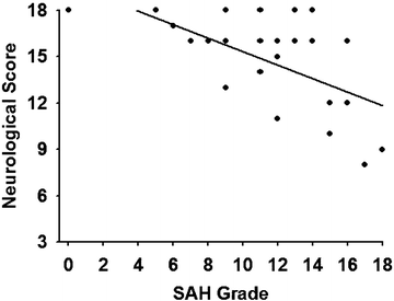
Fig. 4.
Simple regression analysis and Spearman’s correlation coefficient by rank test was performed by Sugawara et al. and found significant correlation between the SAH grade neurological score when using a modified Garcia neurological scale (r = 0.42, p < 0.01) (11, 12). Figure adopted with modification from Sugawara et al. (11).
4.2 Vasospasm
There is a strong correlation between the SAH grade and cerebral vasospasm in the rodent model of endovascular perforation model of SAH. Methods of quantifying vasospasm are provided in other section of this book. Below are data showing the correlations between intracranial IC diameter, perimeter, and wall thickness, which are commonly used histological measurement used to quantify cerebral vasospasm in rodents (Fig. 5).
 < div class='tao-gold-member'>


Only gold members can continue reading. Log In or Register to continue
Stay updated, free articles. Join our Telegram channel
Full access? Get Clinical Tree
