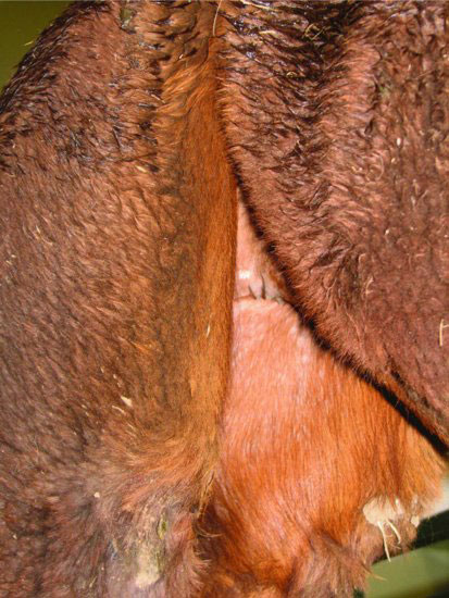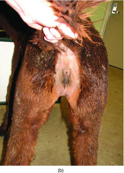
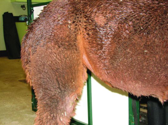
Figure 28.2 Thin fiber coat is seen in the axilla. This is the indicated site for tuberculin skin testing in camelids.
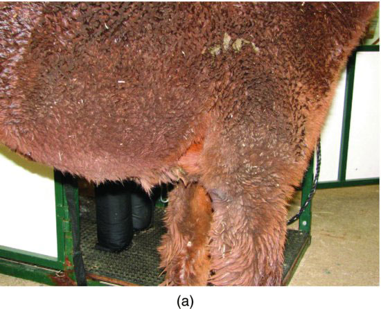
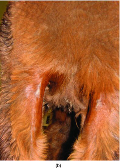
Figure 28.3 Normal thin fiber distribution along ventral abdomen.
Unique integument characteristics should be recognized. The skin is relatively thick, especially in the cervical region of intact males, as well as tense and nonpliable. (See Figure 28.5.) Camelids have functional sweat and sebaceous glands that can initiate abnormal growths in addition to their normal thermoregulatory functions. They also possess normal glandular structures seen at both the medial and lateral metatarsal regions, which are long, thick, scaly skinned areas, and should not be confused with “equine chestnuts” or pathology. (See Figure 28.6.) Also note a similar appearance of the dorsal interdigital space; these are interdigital glands. (See Figure 28.7.) Unlike cloven-hooved species where interdigital pododermatitis commonly affects the interdigital cleft of the hoof, camelids are affected by an infectious pododermatitis that will commonly cause a central, circular, necrotic lesion in the bottom of the foot pad. Generalized crusty, thickened skin may be associated with other dietary deficiencies, such as a zinc responsive dermatosis, immune-mediated or infectious dermatitis and others (VanSaun et al. 2006; Lamm et al. 2009).
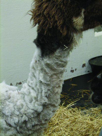
Figure 28.5 Thick cervical skin is expected in camelids, especially intact males. Sometimes venous cutdown is necessary for jugular catheterization.
Stay updated, free articles. Join our Telegram channel

Full access? Get Clinical Tree


