Chapter 114 Amputation of the Digit
ANATOMY
Distal Phalanx (Cats)
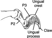
• The proximal end of the third or distal phalanx (P3) is concave and has a dorsal ungual crest (Fig. 114-1).
• The stratum germinativum contains the germinal cells of the claw and extends into the ungual crest.
Dewclaw (Dogs)
• The dewclaw is the medial or first digit of the rear limb in the dog. The first phalanx and P2 are often missing, and P3 and the claw are attached only by skin and fibrous tissue.
• In some breeds, there are two dewclaws. Double dewclaws are breed standard specific in Great Pyrenees and briards and should not be removed.
• The dewclaw articulates with metatarsal bone I, which is often small and may be fused to tarsal bone I.
Digits (Dogs and Cats)
• The digits consist of three phalanges (P1, P2, and P3) and a nail or claw. The first digit in the front foot does not have a middle phalanx (P2); the first digit in the rear foot is properly termed the dewclaw.
• The proximal interphalangeal (PIP) and distal interphalangeal (DIP) joints are connected axially and abaxially by collateral ligaments. The DIP joint is also connected by a dorsal elastic ligament that spans from the dorsal aspect of P2 to the ungual crest of P3.
• The superficial digital flexor tendon attaches to the proximal (palmar or plantar) end of P2; the deep digital flexor attaches to a rounded tubercle on the palmar or plantar aspect of P3; the lateral and common digital extensor tendons attach to the extensor processes of P3.
• A digital pad is located on the palmar and plantar aspect of the distal interphalangeal joint of each digit, except for the first one. A single large metacarpal or metatarsal pad is located on the palmar and plantar aspects, respectively, of the metacarpophalangeal and metatarsophalangeal joints.
• The blood supply is via dorsal and palmar (or plantar) proper digital arteries; venous drainage is via dorsal and palmar (or plantar) proper digital veins.
• The nerve supply to the digits is the dorsal and palmar (or plantar) dorsal digital and dorsal proper digital nerves. Of more functional significance are the nerves that innervate the foot as a whole, the superficial radial, ulnar, and median in the front foot and the tibial, peroneal, and saphenous in the rear foot. Perform nerve blocks with long-acting local anesthetics whenever foot surgery is performed (see Chapter 6).
ONYCHECTOMY (CATS)
Preoperative Considerations
• Many owners are unaware of what a declaw procedure involves. An accurate and frank discussion of the procedure, with anatomic charts, often eliminates confusion and misinformation about the procedure and allows the owner to make an informed decision.
• Inform owners that declawing removes, to varying degrees, the cat’s ability to escape or defend itself. This is especially true when all four feet are declawed. Therefore, most cats should be kept indoors as house pets after declawing. Behavioral issues may develop in multi-cat households when some cats are declawed and others are not.
• It is seldom necessary to remove the claws of the rear limbs. If the owner desires declawing of the rear feet, the consequences should be frankly discussed and other options explored prior to performing the procedure.
• Alternatives to declawing include simple nail trimming on a regular basis, behavioral modification, vinyl caps, and deep digital flexor tenectomy.
Surgical Procedure
Equipment
• Sterile guillotine-type nail trimmers (e.g., Resco); reserve a clean, sharp trimmer for declaws only and replace the blade periodically as it dulls
General Technique
3. Place a tourniquet below the elbow. Exsanguinate the limb to the level of the tourniquet with an Esmarch bandage or hands prior to tightening or inflating the tourniquet. A Penrose drain and Kelly forceps (with tips curving away from the limb if using curved forceps) work reasonably well. Do not use rubber bands or black rubber tourniquets. Use of a CO2 laser may eliminate the need for a tourniquet.
Scalpel Technique
1. Extend the claw by grasping the claw with Kocher forceps. Rotate the forceps and claw downward while tractioning the limb towards you.
2. Hold the scalpel like a paintbrush. Orient the blade transversely to the digit. Beginning at the junction of skin and claw, push the skin proximally, tensing the skin over the DIP joint, and press the scalpel blade downward to cut through the skin, dorsal elastic ligament, digital extensors, and joint capsule.
Stay updated, free articles. Join our Telegram channel

Full access? Get Clinical Tree


