27 Alteration of eyelid position and movement
PRESENTING SIGNS
• Ptosis: half-closed eyes, droopy eyelid(s). The contralateral eye may appear exophthalmic by comparison.
• Narrower or wider palpebral fissure: the eye appears smaller, recessed, sunken, or bigger, bulging or enlarged.
Innervation of the palpebral fissure
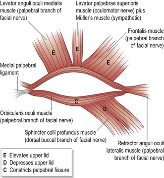
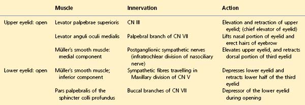
The size of the palpebral fissure is chiefly controlled by CN III and sympathetic neurons which elevate the upper eyelid (Fig. 27.1). Failure of one or both causes a drooping of the upper lid called ptosis (Table 27.1).
Eye closure is achieved by contraction of the orbicularis oculi muscle, innervated by the facial nerve (CN VII) (Table 27.2).
Table 27.2 Closing the palpebral fissure
| Muscle | Innervation | Action |
|---|---|---|
| Orbicularis oculi | Branch of CN VII | Sphincter-like closure of palpebral fissure. Depresses the eyebrow |
| Retractor anguli oculi lateralis | Branch of CN VII | Draws lateral palpebral angle caudally |


