CHAPTER 15 African Horse Sickness
African horse sickness (AHS) is a noncontagious, infectious, insect-borne disease of equids caused by African horse sickness virus (AHSV). In horses the course of the disease is usually peracute to acute, and more than 90% of immunologically naive animals die. Clinically, AHS is characterized by pyrexia; edema of the lungs, pleura, and subcutaneous tissues; and hemorrhages on the serosal surfaces of internal organs. Mules are less susceptible than horses and donkeys, and zebras rarely show clinical signs of disease.
An Arabic document reports the first known historical reference to a disease resembling AHS occurring in Yemen in 1327.1 Father Monclaro’s report of the travels of Francisco Baro in East Africa in 1569 also reports AHS affecting horses imported from India.1,2 Although neither horses nor donkeys were indigenous to southern Africa, they were introduced shortly after the arrival of the first settlers of the Dutch East India Company in the Cape of Good Hope in 1652.1 Records of the Dutch East India Company make frequent reference to “perreziekte” or “pardeziekte” in the Cape of Good Hope.2 In 1719, almost 1700 horses died from AHS in the Cape. During their exploration and expansion into the interior of southern Africa, the Voortrekkers reported severe losses among their horses.1 Exploration of southern, central, and East Africa by Livingstone was complicated by his inability to use horses on some of his journeys.2 Although horses die as a result of AHS every year in southern Africa, major epizootics before the 1950s occurred about every 20 to 30 years. Severe losses were reported in 1780, 1801, 1839, 1855, 1862, 1891, 1914, 1918, 1923, 1940, 1946, and 1953.1 The 1854-55 epizootic was the most severe, with almost 70,000 horses dying, representing more than 40% of the horse population of the Cape of Good Hope.3
Initially, AHS was confused with anthrax and piroplasmosis. In the early 1900s, M’Fadyean,4 Theiler, Nocard, and Sieber1 all succeeded in transmitting the disease with a bacteria-free filtrate of blood from infected horses, confirming that the disease was caused by a virus. The pioneering research of Sir Arnold Theiler, who founded the Veterinary Research Institute at Onderstepoort in 1908, revealed the plurality of “immunologically distinct strains” of AHSV because immunity acquired against one “strain” did not always afford protection against infection by “heterologous strains.”2,5–8 Alexander9,10 showed that viscerotropic isolates of AHSV became neurotropic but did not lose their immunogenicity after serial intracerebral passage in mice. This work led to the development of the first polyvalent vaccine against AHS in the 1930s.11,12 Although Pitchford and Theiler13 proposed in 1903 that AHS may be transmitted by biting insects, it was not until 1944 that Du Toit14 confirmed that Culicoides species were probably vectors of both AHS and bluetongue viruses.
In endemic areas, severe losses caused by AHS have ceased since the development of polyvalent vaccine. However, the occurrence of epizootics in countries outside the endemic regions in Africa15–17 serve as a warning that AHS may spread to areas traditionally free of the disease. AHS is one of the important diseases to consider when moving equids internationally, but movement can be accomplished safely by following appropriate quarantine and testing procedures.18,19
ETIOLOGY
AHSV is a member of the genus Orbivirus in the family Reoviridae and as such is morphologically similar to other orbiviruses, such as bluetongue virus (BTV) of ruminants and equine encephalosis virus (EEV; see Chapter 26).20,21 The virion is not enveloped and is about 70 nm in diameter. It consists of a two-layered icosahedral capsid composed of 32 capsomeres.22 The genome comprises 10 double-stranded ribonucleic acid (RNA) segments, each of which encodes at least one polypeptide.23 The core particle comprises two major proteins, VP3 and VP7, which are highly conserved among the nine AHSV serotypes, and three minor proteins, VP1, VP4, and VP6.22,24 Together these proteins make up the group-specific epitopes.25 The core particle is surrounded by the outer capsid, which is composed of two proteins, VP2 and VP5. VP2 is the protein responsible for antigenic variation.26–28 At least three nonstructural proteins have been identified in infected cells (NS1, NS2, and NS3/3a).29–31
Nine antigenically distinct serotypes have been described.32,33 Although there may be some cross-relatedness between the serotypes, there is no field evidence of any intratypic variation.32,33 All nine serotypes have been documented in eastern34 and southern32,33,35 Africa, whereas serotype 9 is more widespread and appears to predominate in the northern parts of sub-Saharan Africa.36,37
EPIDEMIOLOGY
AHSV is biologically transmitted by Culicoides spp., of which C. imicola and C. bolitinos have been shown to play an important role in Africa.38,39 The disease therefore has a seasonal occurrence, and its prevalence is influenced by climatic and other conditions that favor the breeding of Culicoides spp. Culicoides variipennis, a midge prevalent in the United States but not present in Africa, has been shown to transmit AHSV under laboratory conditions.40 Although other insects have been suggested as possible vectors of AHSV, none has been shown to play a role under natural conditions. Biting flies may play a minor role in the mechanical transmission of AHS; however, because the viremia in horses is relatively low and AHSV is susceptible to desiccation and high temperature, this method of transmission is inefficient. AHSV can be transmitted between horses by parenteral inoculation of infective blood or organ suspensions, and it is more readily transmitted by the intravenous than by the subcutaneous route.1
A continuous transmission cycle of AHSV between Culicoides midges and zebras was shown to exist in the Kruger National Park in South Africa.41 Under such circumstances, a sufficiently large zebra population can act as a reservoir for the virus.42–44 Donkeys may play a similar role in parts of Africa with large donkey populations.45 In view of the high mortality in horses, this species is regarded as an accidental or indicator host. Animals that have been infected with AHSV do not remain carriers of the virus, which explains the failure of the disease to become established outside tropical Africa, despite the occurrence of many outbreaks outside endemic areas.1
AHS is endemic in eastern and central Africa2 and spreads regularly to southern Africa. In endemic areas, different serotypes of AHS may be active simultaneously, but one serotype usually dominates during a particular season. The disease is also reported from time to time in countries in North Africa, from where it has occasionally extended into the Middle East and Spain.46,47 However, its intrusion into North Africa and countries around the Mediterranean and in Asia is impeded by the Sahara desert.48 AHS has not been recorded in Madagascar or Mauritius.
AHS was recorded in Egypt in 1928, 1943, 1953, 1958, and 1971;49 in Yemen in 1930; and in Palestine, Syria, Lebanon, and Jordan in 1944.46,50–52 In 1959, AHS serotype 9 occurred in the southeastern regions of Iran. This was followed by outbreaks during 1960 in Cyprus, Iraq, Syria, Lebanon, and Jordan, as well as in Afghanistan, Pakistan, India, and Turkey. Between 1959 and 1961, this region lost more than 300,000 equids.52,53 In 1965, AHS occurred in Libya, Tunisia, Algeria, and Morocco and subsequently spread to Spain in 1966.54 Between 1987 and 1990, AHS serotype 4 occurred in Spain, with the source of infection being zebra (Equus burchelli) imported from Namibia.16,17 AHS was also confirmed in southern Portugal in 1989 and Morocco between 1989 and 1991, with these outbreaks being extensions of the outbreak in Spain.16,55 In 1989 an outbreak of AHS serotype 9 occurred in Saudi Arabia.15 AHS was also reported in Saudi Arabia and Yemen in 1997 and on the Cape Verde Islands in 1999.
AHS can be distributed over great distances if equids incubating the disease are translocated by land, sea, or air.17,56 Outbreaks have also been reported to result from wind-borne spread of infected vectors.57
AHS is not endemic in parts of South Africa, but each year the disease appears in the northeastern part of the country, occasionally during December but usually in January, from where it spreads southward. The extent of the southerly spread is influenced by the extent of favorable climatic conditions for the breeding of Culicoides midges.48 Early and heavy rains followed by warm, dry spells favor the occurrence of epizootics. Although parts of the inland plateau of South Africa and most of the Cape Province are usually free of AHS, the disease has sometimes extended into these areas and caused serious losses. Significant losses were reported at Belfast, a town situated about 2100 meters (8000 feet) above sea level during the severe epizootic of 1923.58 The first cases of AHS usually occur at the beginning of February, but the most serious outbreaks usually occur in March and April. Following the first frosts, which usually occur at the end of April or in May, the disease disappears abruptly. However, in the northeastern parts of South Africa, where the occurrence of frost is less common, deaths may continue to occur into May and June.1 In recent years the southerly spread of AHS has been less extensive, probably as a result of the widespread use of a more effective vaccine, which became available in 1974.48 Approximately 300,000 doses of polyvalent AHS vaccine are sold annually by Onderstepoort Biological Products. It is speculated that immunization of horses in these regions establishes a fairly effective “immune barrier” that seems to impede the southerly spread of the disease.48 Outbreaks of AHS associated with the introduction of infected animals have been reported in the Cape Peninsula in 1967,48 1990,59 1999,60 and 2004.
AHS affects primarily equine animals. Horses are most susceptible to the disease (mortality of 70%-95%), but mules are less so (mortality of 50%-70%). Most infections of donkeys and zebras are subclinical.2,46 Generally, horses of all breeds are equally susceptible to AHS, but variation in susceptibility to the same virus in individual horses has been reported.2 Some indigenous horses in North and West Africa, which descend from animals that have been present there since at least 2000 bc, have apparently acquired natural resistance to AHS.61 Foals born to immune mares acquire passive immunity by the ingestion of colostrum.62 This passive immunity progressively declines and is completely lost after about 4 to 6 months. Donkeys in the Middle East appear to be more susceptible to AHS (mortality of 3%-10%) than southern African donkeys.46 Zebras are highly resistant to AHS and only show a mild fever after experimental infection.63
Dogs are the only other species that contract a highly fatal form of AHS.64–67 All reported clinical cases in dogs have resulted from the ingestion of infected carcass material from horses that have died from AHS.68,69 AHSV serotype 9 has been isolated from the blood of stray dogs in Egypt,49 and antibodies to AHSV have been detected in the sera of dogs in India70 and South Africa.71 However, it is doubtful that dogs play any role in the spread or maintenance of AHSV, because Culicoides spp. do not readily feed on them.71 Besides zebras,63,72–74 no other wildlife or domestic ruminants have been shown to play a significant role in the epidemiology of AHS.
PATHOGENESIS
After infection, initial multiplication of AHSV occurs in the regional lymph nodes and is followed by a primary viremia,75 with subsequent dissemination to endothelial cells of target organs.76 Effusions into body cavities and edematous changes of various tissues, as well as serosal and visceral hemorrhages, are consistent with endothelial cell damage. In experimental cases of AHS, high virus concentrations are found in the spleen, lungs, cecum, pharynx, choroid plexus, and most lymph nodes by the second day after inoculation. This precedes the onset of fever or detectable viremia. By the third day after inoculation, virus is present in most organs. Virus multiplication at these sites gives rise to a secondary viremia of variable duration. In horses the viremia is generally not higher than 105 TCID50/mL and lasts 4 to 8 days but does not exceed 21 days. In donkeys and zebras the viremia is lower but may last as long as 4 weeks.75 In zebras, viremia has been reported in the presence of circulating antibodies.63
The factors determining the course and severity of infection are not well understood. Small plaque variants produce severe clinical reactions, whereas large plaque variants of AHSV are less pathogenic.75 Fully susceptible horses, such as foals that have lost their colostral immunity or horses that have never been exposed to the AHSV, usually develop the peracute “pulmonary” form of AHS. Exercise during the febrile stage of the disease may also precipitate this form of AHS.50,75 During the 1959 Middle East epizootic of AHS caused by serotype 9, severe myocardial lesions with extensive areas of degeneration and necrosis of myocytes, accompanied by a marked inflammatory response, were described in fully susceptible horses, particularly those with the “cardiac” form of the disease.77 In this form of AHS, heart failure is attributed to hydropericardium75,78 and myocardial damage.77 However, most natural cases of AHS are of the “mixed” form, with evidence of both pulmonary and cardiac compromise.47,75,77
CLINICAL FINDINGS
The clinical findings of natural and experimental cases of AHS have been described.17,77,79–81 In experimental cases the incubation period is usually between 5 and 7 days, but possibly as short as 2 days and rarely as long as 10 days. The duration of the incubation period depends on the virulence of the virus and the dose of virus received.
“Dunkop” or “Pulmonary” Form
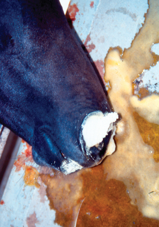
The incubation period for pulmonary AHS is short, usually 3 to 4 days, and is followed by a rapid rise in temperature over 1 or 2 days, with the body temperature reaching 104° to 106° F (40°-41° C). The dunkop form is characterized by marked and rapidly progressive respiratory failure, and the respiratory rate may exceed 50 breaths per minute. The animal tends to stand with its forelegs spread apart, its head extended, and the nostrils dilated. Expiration is frequently forced, with the presence of abdominal heave lines. Profuse sweating is common, and paroxysmal coughing may be observed terminally, often with frothy, serofibrinous fluid exuding from the nostrils (Fig. 15-1). The onset of dyspnea is usually very sudden, and death occurs within 30 minutes to a few hours of its appearance. Sometimes an apparently healthy horse at work becomes listless, suddenly severely dyspneic, and dies shortly thereafter. Initially, the appetite of affected animals remains good despite the high fever and respiratory distress. The prognosis for horses with the dunkop form is extremely poor (<5% recover). If animals recover, the fever subsides gradually, but the breathing remains labored for several days.
“Dikkop” or “Cardiac” Form
The incubation period in the “dikkop” or “cardiac” form of AHS is longer than the “dunkop” (pulmonary) form, usually 5 to 7 days, followed by a fever of 102° to 106° F (39°-41° C) that persists for 3 to 4 days. The more typical clinical signs do not appear until the fever has begun to decline. At first the supraorbital fossae fill as the underlying adipose tissue becomes edematous and raises the skin well above the level of the zygomatic arch (Figs. 15-2 and 15-3). The edema can later extend to the conjunctiva (Fig. 15-4), lips, cheeks, tongue, intermandibular space, and laryngeal region and may extend a variable distance down the neck toward the chest, often obliterating the jugular groove. As the swellings increase, dyspnea and cyanosis may supervene. However, ventral edema and edema of the lower limbs are not observed. Unfavorable prognostic signs include petechial hemorrhages on the conjunctivae and on the ventral surface of the tongue; if they occur, these hemorrhages become evident shortly before death. Some animals may show signs of severe colic, repeatedly lie down, are restless when standing, and frequently paw the ground.
Stay updated, free articles. Join our Telegram channel

Full access? Get Clinical Tree


