9: DIAGNOSTIC IMAGING OF THE HEAD, WITHERS, MANUS AND PES

9 DIAGNOSTIC IMAGING OF THE HEAD, WITHERS, MANUS AND PES


Clinical considerations for diagnostic imaging


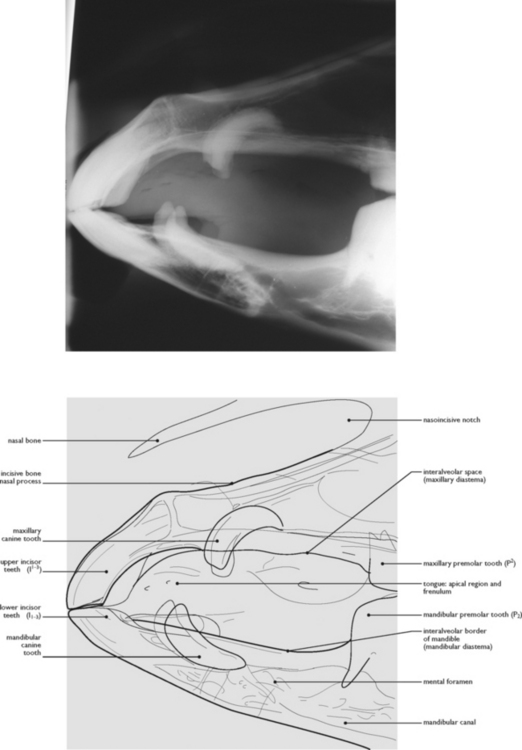
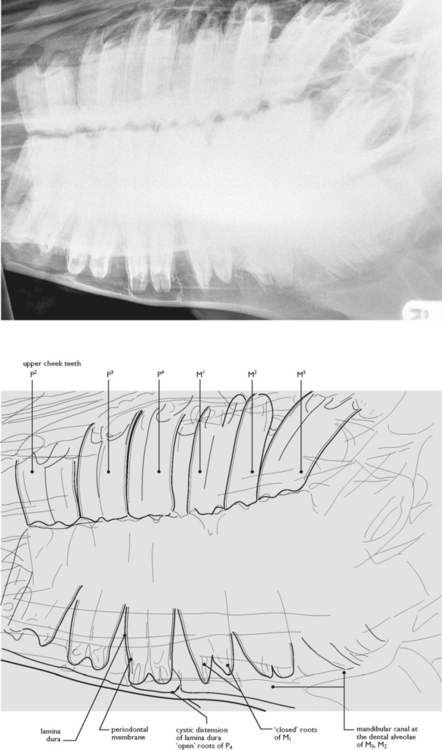
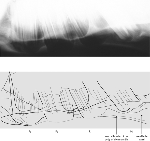
Radiography is a very useful diagnostic tool in equine medicine, particularly in the skull and the distal limbs, although higher-powered equipment will also enable images of the spine, proximal limbs and pelvis to be made. However, in order to be able to interpret radiological abnormalities appropriately, knowledge of normal radiological anatomy must be gained. In general, the distal limb of the equine animal is not too complex. In contrast, the head is extremely complex and so it is of paramount importance to have an atlas of normal radiographic anatomy, or a normal radiograph, available for comparison when attempting interpretation of this area.


The commonest clinical indication for radiography of the limbs is lameness, and the commonest causes of lameness in equine animals are tendon or ligament problems (tendinitis or desmitis), inflammatory conditions (such as laminitis affecting the laminae of the hoof) and trauma (soft tissue injuries and fractures). Although radiography will not demonstrate primary changes in the soft tissue structures, it may demonstrate secondary effects on the bones, such as degenerative joint disease or sclerosis (when the bone becomes dense and hard).


At least two views of the affected area should be taken (orthogonal projections) and, in the case of the limbs, the contralateral limb may also be imaged, to provide a control for comparison. In the distal limb, it is routine to take at least two additional oblique projections of the region of interest in order to assess as much of the region as possible.


Advanced cross-sectional imaging modalities, such as computed tomography (CT), allow appreciation of anatomy without superimposition from overlying structures. CT is of particular value in assessing bony structures, so is particularly useful in demonstrating incomplete fractures and for characterising comminuted fractures (in which the bone has been fragmented into multiple pieces). The lack of superimposition provided by CT is particularly useful in the head, although it is also used to demonstrate change in the limb bones. If appropriate equipment is available, CT may be performed in the standing animal, obviating the need for general anaesthesia.


The presence of cartilaginous epiphyseal (or growth) plates must be remembered when interpreting radiographs of the limbs in young horses. The following times of epiphyseal fusion have been determined radiographically:


Epiphyseal fusion is a gradual process. The epiphyseal growth plate stops producing new metaphyseal bone and becomes itself converted to bone, uniting the epiphysis with the shaft. The process can be traced radiographically or histologically until fusion is complete. Gross anatomical fusion, however, is ‘complete fusion’, when a standard maceration technique fails to separate the epiphysis from the shaft. For the horse, the results of some investigations by these three methods of study are summarized by Getty in Sisson & Grossman (1975, pp 272 & 298).


< div class='tao-gold-member'>

Only gold members can continue reading. Log In or Register to continue

Stay updated, free articles. Join our Telegram channel

Jul 8, 2016 | Posted by in SUGERY, ORTHOPEDICS & ANESTHESIA | Comments Off on 9: DIAGNOSTIC IMAGING OF THE HEAD, WITHERS, MANUS AND PES

Full access? Get Clinical Tree

Get Clinical Tree app for offline access