8: THE PELVIS (INCLUDING THE SPINE)

8 THE PELVIS (INCLUDING THE SPINE)


Clinical importance of the pelvis


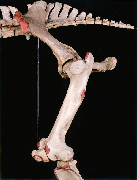
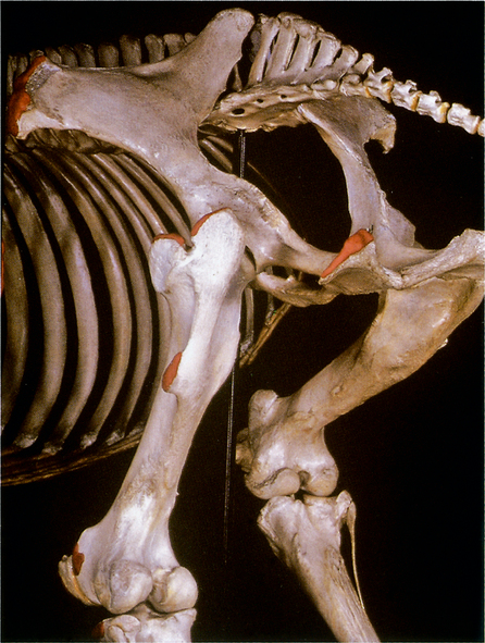
Lameness associated with the pelvis can be investigated by clinical examination, ultrasonography, scintigraphy, radiology in the young horse, local anaesthesia and nerve blocks. The pelvis has a very important function in providing the link between the spine (for support) and the hindlimb (for propulsion). At times the pelvis may be carrying the whole weight of the horse. It transfers the propulsive effort but, because of the strong ligamentous and musculature attachments, has very limited movement itself. It is continuously subjected to non-compressive forces. It could be said that the sacro-iliac ligaments and joint are subjected to the most disruptive forces in the locomotory system. The pelvis gives passage, through the greater sciatic foramen, to the cranial gluteal artery and vein and the gluteal nerves; also to the largest single nerve in the equine body, the sciatic nerve. In addition, the obturator foramen gives passage to the obturator nerves and vessels.


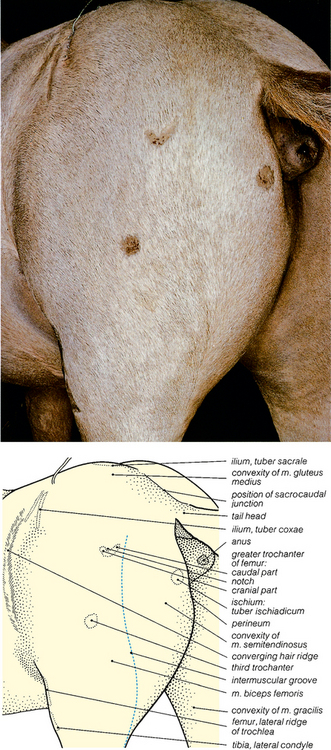
Fractures of the pelvis are relatively rare, as the pelvis is protected by such strong muscle groups. They do, however, occur in traumatic accidents or may present as stress fractures. Often the horse with a pelvic injury may be in shock and may be very reluctant to bear weight or move. There may be fractures of the wing or the shaft of the ilium, acetabulum, pubic bone or ischium. The most common fracture is probably that of the tuber coxae. Pelvic fractures present as a sudden onset of very acute unilateral or bilateral lameness, often following exercise and trauma. The sacral or ischiatic tuberosities often appear uneven in height when viewed from behind.


The sacro-iliac joint is clinically the most important locomotory structure in the pelvis. It is a diarthrodial joint; it has a joint capsule, is thin and tight, with a total capacity of only 1–2 ml. There is hyaline cartilage on the sacral joint surface and fibro-cartilage on the ilial surface. The shape of the articulation varies with age and between individuals so there is considerable variation. The joint is stabilized by three pairs of sacro-iliac ligaments. The dorsal sacro-iliac ligament is divided into dorsal and lateral parts. There is an interosseus ligament and also a ventral ligament. Hunters and jumpers are particularly affected by damage to these ligaments. Which results in a sore back or persistent hindlimb lameness. The injuries fall into two groups. Firstly, there are acute injuries, desmitis, and luxation/subluxation of the joint. Secondly, there is chronic injury in the form of sacro-iliac osteoarthritis or desmitis or both. Both are seen as hindlimb lameness and/or poor performance. The acute injuries result from a fall, from slipping, or from overwork. Which often causes overstretching of the dorsal and ventral ligaments. Sometimes, if the sacro-iliac joint is particularly unstable, it is possible to see movement of the tuber sacrale when the affected horse moves forward at a walk. Chronic sacro-iliac injury results from over-use, trauma, and the saddle. It has a high incidence of 8–15% in racehorses. There is usually a history of poor performance, resisting jumping, lack of hindlimb impulsion, behavioural changes and back soreness. Examination of the joint by ultrasonography often reveals degenerative changes in the joint. Anaesthesia of the joint will confirm the seat of lameness. For the right sacro-iliac joint, a site 2 cm cranial to the left tuber sacrale, at a 45–60 degree angle to the joint and slightly caudal towards the right greater trochanter of the femur, along the medial aspect of the right ilial wing, will reach the joint and vice versa for the left joint. Nerve damage within the pelvic cavity may affect the obturator nerve, which is sometimes damaged in foaling. This nerve innervates the adductor, gracilis, and pectineus muscles. Loss of adduction of the hindlimb results in a wide stance, with inability to keep the limb under the body. Trauma to the cranial and caudal gluteal nerves occurs sometimes, with a resultant atrophy of the gluteo-biceps (biceps femoris) muscle.


‘Ruptures’ occur when a part of the body is displaced through a break in bodily structure. (‘Hernias’ are displacements through natural bodily openings.) Ruptures are often associated with kicks, being stuck half over fences, and road traffic accidents. Damage to the prepubic tendon, followed by rupture, is occasionally described in association with the presence of twin foals, hydro-allantois or giant fetal oversize. Rupture of the abdominal muscles may occur following trauma. Inguinal or scrotal hernias may occur in colts (usually congenital), stallions, or (rarely) mares or geldings. A segment of small intestine, or part of the great omentum, or both, enter the vaginal process through the vaginal ring. These parts may remain close to the superficial inguinal ring (inguinal hernia) or may enter the scrotum (scrotal hernia). In scrotal hernia, the hernial sac comprises the scrotal skin, tunica dartos, and the tunica vaginalis of the vaginal process, and also contains the testis, and epididymis. If the blood supply to the contents of the hernial sac is affected (strangulated hernia), surgery is urgently needed. Surgical treatment usually involves replacing the abdominal contents by twisting the vaginal process. The twisted neck of the process is then sutured to the edges of the superficial inguinal ring (abdominal and pelvic tendons of the external abdominal oblique muscle). The vaginal tunic is incised and the testis and epidymis are removed. Femoral hernia (or ?rupture) occurs through the femoral ring, alongside the femoral artery, and is rare, but (as in the human cases) may be more common in females than in males.


Examination of the pelvis by rectal examination is not so easy in the horse as in the ox because the rectum is friable and easily torn. It can be used to evaluate inguinal hernias and cryptorchidism (undescended testis). If a stallion has colic, it can be used to investigate whether there is an incarceration of the intestine in the inguinal canal. The superficial inguinal ring is located 6–8 cm cranial to the iliopectineal eminence of the pubis and 10–12 cm lateral to the midline. The perineal region is a favoured area for the occurrence of melanoma in grey horses.


The male and female reproductive tracts are associated with the pelvic region and will be discussed here. The intra-abdominal features of both female and male systems can be viewed by laparoscopy. The horse is a long-day breeder and is seasonally polyoestrus. Examination of the external and internal genital tract of the mare is an important aspect of stud practice. Vaginal examination using a speculum, and digital examination, are possible. Vulval bleeding occurs occasionally. One of the more unusual conditions is pneumo-vagina (wind-sucking) which constitutes a risk for subsequent bacterial infection. There may be fibroids and sacculations of the uterus. Trans-rectal ultrasonography is very useful in pregnancy, to assess the ovarian follicles and corpora lutea, the presence of small or hypoplastic ovaries, the presence and size of follicles, and the state of the uterus. It is particularly important to detect endometritis or chronic endometriosis. The most important diagnosis is Contagious Equine ‘Metritis’ caused by Tayloriella equigenitalis which is the most important infectious genital disease of the mare and stallion. Endometrial cysts, peri-glandular fibrosis, and cystic glandular distension, may also be revealed. Mucometra (mucus accumulations in the uterus) also occurs occasionally. Enlarged ovaries may indicate tumours such as granulosa-cell tumours. Endoscopy can be used to collect microbiological samples and samples for endometrial biopsy or cytology.


Uterine torsion is not uncommon as a cause of colic in the late term mare (usually at around 8 months). It may be 180–540 degrees torsion, cranial to the cervix and vagina, and rupture of the uterus may result from the torsion.


Placentitis is an unusual occurrence. It may be necessary to carry out ovariectomy because of abnormal behaviour, to curtail reproductive activity, or for pathological conditions such as granulosa-cell tumours or haematomas in the ovary. The operation can be carried out through the flank or by ventral midline, oblique paramedian or vaginal approaches. Inflammation of the Fallopian tubes (oophritis) may also require their removal.


Pregnancy failure in the mare is not uncommon as a result of embryonic death or abortion. Pregnancy diagnosis in the mare is possible per rectum and is accurate after about 28 days and optimal around day 42. Trans-rectal ultra-sonography will give results from day 10 to term. Usually the first scan is 14–15 days. To detect twins before implantation, scan at 16 days. The heart beat should be visible at 26–28 days. Transabdominal ultra-sonography can be used from day 80 to term. It is possible during pregnancy to have umbilical cord abnormalities, placental insufficiencies, pregnancy in the uterine body, insufficiency of hormones from the corpora lutea and, the worst possibility of all, uterine torsion. Prolonged gestation is rare, with only 1% exceeding 370 days. Premature udder development occurs occasionally. There are several causes of abortion including bacterial, fungal, and viral agents. In the latter group, equine infectious anaemia, equine herpes virus 1 and equine viral arteritis, are the most important.


Parturition is a time when intervention may be necessary. Caesarean section is essential if second stage labour is delayed, but it is rare to get a live foal delivered by this operation. From the mare’s point of view it is preferable to perform an embryotomy. Parturition occurs in 3 stages. The first stage, which lasts about one hour, is rupture of the fetal membranes; the second stage lasts about 30 minutes and the foal is born. Stage three relates to expulsion of the foetal membranes and lasts about 1 hour. Uterine involution takes about 6–10 days and the foaling heat occurs 7–9 days after foaling. Post-partum complications include retained placenta, haemorrhage, recto-vaginal fistula, perineal lacerations, uterine rupture, endometrial haemorrhage, uterine prolapse, and endometritis. Dystocia (difficult parturition) is a rare event with only about 4% of foalings having difficulty. It may be followed by acute septic metritis or necrotic vaginitis; rupture of the internal iliac artery rupture has been known after a long period of dystocia. Interventions which may prove necessary include retropulsion, forced extraction, amputation of fetal parts (foetotomy), or Caesarean section. The latter operation requires mid-line laparotomy with an incision along the greater curvature of the uterus.


Examination of the stallion for soundness requires examination of the musculo-skeletal, respiratory, and cardiovascular systems, as well as the reproductive tract and semen evaluation. The semen should have a high progressive motility, a pH of 7.2–7.6, a volume of around 70 ml (quite variable but not too important), a concentration of 8 × 10⁶ and, in terms of morphology, at least 60% should be normal and 10% still motile after 6 hours. If a second sample is taken on the same day, the values should be at least 50% of the first sample. There are ejaculatory dysfunctions, some of which may have an anatomical basis, or an abnormality of the locomotory system may make it difficult to mount. The external genitalia have to be examined. Penile trauma is quite common and includes abrasions, lacerations and rupture of the corpus cavernosum penis. The penis can be affected by a variety of conditions including inability to retract the organ (due to inanition or debility after breeding), haematomas, coital exanthema, inflammation (balanitis) and, rarely, squamous cell carcinoma and papilloma. Removal of these tumours may require surgery. Contraction of the preputial ring can also occur following injury and may prevent extrusion of the penis (acquired phimosis). The prepuce can be an important source of bacterial pathogens, particularly for the mare at mating. Examination of the scrotum may reveal acquired swellings, acute trauma, scrotal lacerations, testicular torsions or even hydrocoele (fluid within the vaginal process).


The scrotum is an area that is often involved in trauma, so may show signs of scars on its surface. Torsion of the spermatic cords is an extremely rare event. Congenital abnormalities of the scrotum may be a result of male pseudo-hermaphroditism or the testicular feminization syndrome. The testes should be palpated and their size and consistency are related to the use of the stallion and his daily sperm output. The left testis is often larger and more pendulous than the right. Abnormalities may include hypoplasia (revealed in the young horse), degeneration of the testes following trauma, inflammation (orchitis) which may follow trauma or bacterial infection or both, and neoplasia in the form of a seminoma. It may also be possible to palpate the epididymis (which is on the dorso-lateral border of the testis) for abnormalities. The tail of the epididymis is at the caudal pole of the testis. The internal genitalia may be palpated per rectum.


Castration of the stallion (also known as gelding) is one of the most common operations carried out in equine practice and is usually carried out to prevent male behaviour, rather than for medical or surgical reasons. The exception to this is for the horse that has an undescended testis (cryptorchid). There are open and closed techniques for castration. The operation can be carried out in the field, in the standing position, or in lateral recumbency. The operation can be followed by a variety of complications (infection, haemorrhage, swellings, septicaemia, tetanus, schirrhous cord, hydrocoele, penile prolapse, evisceration, and furunculosis) so care must be taken with hygiene. In the cryptorchid horse (‘rig’) the testis and epididymis fail to descend into the scrotum. In half of these cases, the testis is retained in the abdomen (usually near the deep inguinal ring) and the vaginal process is absent, or short (containing only the tail of the epididymis). In the other cases the testis lies in the inguinal region or canal. It may lie superficial to the superficial inguinal ring of the external abdominal oblique muscle, but it may not be palpable externally. In this position it will be lying between the origin of the lateral scrotal lamina from the dorsolateral edge of the ring (abdominal tendon of the external abdominal oblique muscle) and the origin of the medial scrotal lamina from the yellow elastic lamina medial to the ring. The cryptorchid testis in the inguinal position is usually much smaller than the descended testis, and the scrotum may be poorly developed. In most cases it can be removed by surgery in the inguinal region but some cases require a paramedian abdominal incision.


Clinical importance of the spine


Clinical considerations of the spine involve the regions of the head, neck, thorax, abdomen, and pelvis. Changes in the conformation, musculature and general well-being may affect the spinal column. Clinical examination, lameness tests, regional anaesthesia, diagnostic and contrast radiography and scintigraphy, are widely used in diagnosis. Clinical examination at exercise, either in hand or by lunging, are used in diagnosis. Also, marks may be seen and swellings detected on palpation.


There are only 36 bones in the equine vertebral column (excluding the tail), but there are many ligaments. Some are extremely powerful, such as the nuchal and supra-spinous ligaments, and there are many smaller ligaments such as the interspinous, sacroiliac, sacrotuberous, iliolumbar, intertransverse and dorsal and ventral longitudinal ligaments. Clinical signs that suggest spinal problems include acute or chronic ‘back’ pain, reduced flexibility of the spine, lateral curvature of the spine, asymmetry of the hindquarters, neurological deficits and atrophy of the dorsal muscles and hindquarters.


The diseases and disorders of the spinal column, dorsal musculature, and central nervous system are sometimes extremely difficult to differentiate. Many conditions of the back are diagnosed by elimination of the other possibilities, rather than by the finding of probable diagnostic indicators. ‘Back problems’ often have very indeterminate clinical signs such as poor performance, behavioural or temperamental changes, and difficulty of control when being mounted or ridden. Reluctance to go faster or to jump is also a very common feature. Vertebral trauma occurs quite often when horses fall at speed, when taking jumps, or in serious collisions. Fracture of the spinous processes, vertebral body and neural arches is most likely in the first three thoracic, first three lumbar and T12 vetebrae. If the horse falls over backwards it is more likely to suffer from fractures of the dorsal spinous processes. In many of these cases the horse will remain on the ground or be ‘winded’. Fractures or other injuries of the cervical, thoracic, and lumbar vertebrae are not uncommon in these types of trauma, and may be accompanied by fractures of the long bones and/or accessory carpal bone, making diagnosis even more difficult. A horse pulling up lame is more likely to have problems with the superficial digital flexor tendons, or fracture of the accessory carpal bone or a lateral condyle.


The spine may be subjected to a variety of conditions. Abnormalities of conformation are seen in foals; congenital scoliosis (lateral curvature) is not uncommon; much less common are lordosis (ventral curvature), kyphosis (dorsal curvature) and synostosis (fusion of the vertebrae). Muscular damage affecting the spine includes strains, myopathies, myositis, white muscle disease (vitamin E and selenium deficiency) and external rhabdomyolysis. Ligamentous strains are also very common. Misalignment of the vertebrae leading to pinched nerves is also not uncommon. One of the most common conditions is over-riding or fracture of the spinal processes of the thoracic vertebrae. Spondylitis is inflammation of a vertebra; ossifying spondylitis and discospondylolisthesis are associated with traumatic incidents and are not uncommon.


One of the most common causes of back pain in the horse is the condition known as ‘kissing spines’, in which there is crowding or over-riding of the spinous processes of the vertebrae under the saddle area. It has a high incidence and a very low mortality and is caused by conformational abnormalities. It causes pressure points between the adjacent spines with over-riding. Adventitious bursae develop between the spines in response to the persistent rubbing, and where there is persistent over-riding new bone develops in response. Affected horses buck when ridden, have a very poor hindlimb action, and a poor jumping performance. The rider’s weight is usually carried by T12–T18 and the most likely sites of lesions are in T15–T17.


In older horses there are also conditions associated with loss of strength in the ligaments around the small joints of the vertebrae, causing lesions in the transverse processes and the articular processes of the vertebrae.


In considering the spine as a whole it is important to realise that most of the conditions likely to be a common cause of abnormalities are found in the pelvic region. The sacral vertebrae are an important part of the spinal column and the sacroiliac ligaments can be easily damaged (see Pelvic section). One of the conditions not mentioned in the pelvic section is fracture of the sacrum. In this injury, the peripheral nerves of the hindleg (sciatic, fibular, tibial and obturator) are often involved. The femoral nerve is extremely well protected and less likely to be damaged. Fracture of the sacrum may also cause neuritis of the cauda equina, which affects sacral and coccygeal nerve roots.


The neurological problems of the equine spine can be investigated by radiography, ultrasonography, myelography, and by collection and analysis of cerebrospinal fluid. The fluid is best collected with the horse under a general anaesthetic, in lateral recumbency, with the head flexed to ninety degrees and with a straight spinal column. The needle is inserted in the midline between the cranial borders of the wings of the atlas, to a depth of 5–8 cm. Fluid can also be collected from the lumbo-sacral space. For this the horse, needs to be sedated, restrained and standing level on all four feet. The needle is inserted on a line that joins the cranial borders of the left and right tuber coxae, at the intersection with the midline. One to two cm behind this intersection is a depression; the caudal aspect of the 6th lumbar vertebra lies cranially and the cranial border of the 2nd sacral spinous process lies caudally; the rim of the tuber sacrale lies laterally. It is important to remember that L5 is less prominent than L6 and sometimes difficult to palpate, as is S1 which is heads promiuent than S2. The sub-arachnoid space can then be located at a depth of 12–13 cm.


Infectious disease of the equine spine is not an uncommon phenomenon. In many parts of the world viral encephalitides are common, including Eastern, Western and Venezuelan equine encephalomyelitis. Equine herpes virus 1 and West Nile virus infections may also occur. Various other disorders also affect the central nervous system including equine degenerating myelo-encephalopathy (EDM), verminous encephalitis (associated with aberrant parasites), equine protozoal myelitis, ‘ryegrass staggers’, skeletal mycotoxicoses, bacterial meningitis, botulism, tetanic hypocalcaemia, hyperkalaemic periodic paralysis and fungal meningitis.


Neural disorders of uncertain aetiology include post-endurance-race cerebral syndrome, pareses associated with upper and lower motor neuron diseases, trembling, and ataxia. This latter may be induced by plant poisons and may be seen as a lack of proprioception, and a syndrome involving both the vestibular system and the cerebellum. Neuroses include crib-biting, head-shaking, nodding (may be associated with allergy to inhaled antigens), self-mutilation syndrome and wind-sucking.


Tail damage is usually seen as a tail with ‘kinks’ or with a tendency to be flaccid. However, these signs can be caused by sacral fractures which result in nerve damage and neurogenic atrophy of sacral and coccygeal muscles.


image

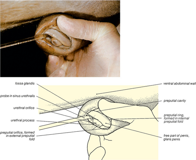
Fig. 8.5 Surface features of the penis and prepuce of the gelding: left lateral view. At birth, the free part of the penis is adherent to the internal lamina of the prepuce (see Fig. 8.47) but this adherence rapidly breaks down (Figs. 8.48 to 8.53) and in geldings and stallions the free part of the penis is separated from the prepuce by the preputial cavity. The urethral process is enclosed by a narrow cavity, the fossa glandis; the sinus urethralis is a dorsal enlargement of this fossa, and its position is indicated by the probe.


image

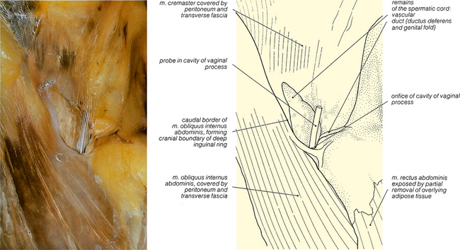
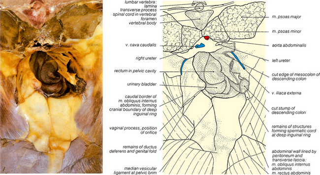
Fig. 8.7 The right inguinal canal of the gelding: cranial view. This is a closer view of a part of the specimen shown in Fig. 8.6, at a slightly later stage in the dissection. After castration, the components of the spermatic cord atrophy. A probe has been inserted into the persistent and still patent orifice leading to the cavity of the remaining stump of the vaginal process.

< div class='tao-gold-member'>

Only gold members can continue reading. Log In or Register to continue

Stay updated, free articles. Join our Telegram channel

Jul 8, 2016 | Posted by in SUGERY, ORTHOPEDICS & ANESTHESIA | Comments Off on 8: THE PELVIS (INCLUDING THE SPINE)

Full access? Get Clinical Tree

Get Clinical Tree app for offline access