7 Recognition and Resuscitation of the Critically Ill Foal
Case 7-1 Foal in Hemodynamic Shock
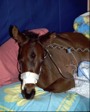
’04 Revival was admitted to the NICU when he was 15 hours old (Figure 7-1). The foal arrived recumbent and required transport to the NICU. As is the standard of practice, vital signs and essential organ function were rapidly evaluated while supportive therapy was initiated. This was done using a team approach. The following tasks were performed simultaneously: intranasal oxygen cannula placed, jugular vein prepared for catheterization, TPR data collected, rapid physical assessment of essential organ function made, arterial sample for blood gas and electrolytes obtained (before intranasal oxygen insufflation initiated as room air baseline), and indirect blood pressures recorded. As the intravenous catheter was placed, blood for culture and laboratory analysis was obtained and intravenous fluid administration was initiated as the catheter was secured. This was accomplished within 10 minutes of arrival.
Initial examination of essential organ function: Oral, nasal, and scleral membranes had mild to moderate large-vessel and small-vessel injection and all had marked icterus. Multiple oral petechiae were present as well as marked lingual erythema. Normal respiratory pattern was present with little respiratory effort and no abnormal lung sounds. Normal cardiac rhythm with a relative bradycardia (90 to 96) was present considering that shock was evident. There was an apparent temperature gradient from periphery to core with ice-cold hooves, cold lower legs, and cool nose, ears, and upper legs. The foal had very weak pulses, poor arterial fill, and poor arterial tone. There was evidence of recent urination. Abdominal auscultation revealed few, quiet borborygmi, but the abdomen was primarily silent. On abdominal palpation, meconium was found in the right dorsal colon but little elsewhere. The foal had a history of fetal diarrhea which appeared to be subsiding at the time of admission. The foal was somnolent with occasional struggling, but the struggling appeared meaningful. The foal was able to be aroused and responsive to stimuli, but was subdued.
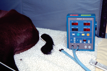
BP is a vital sign that should be measured in all critical neonates (Figure 7-2). Except in cases with severe hypotension and vasoplegia, taking indirect measurements using the oscillometric technique is generally adequate. With this technique, as the pressure in the cuff falls below the systolic pressure, the artery begins to pulsate. These pulsations are transmitted to the cuff, and oscillations are sensed by a transducer. The pressure transmitting maximum oscillations closely correlates to the mean BP. The pressure at which the cuff oscillations begin is recorded as the systolic pressure, and the pressure at which the oscillations stop decreasing is recorded as the diastolic pressure. The instrument eliminates internal artifacts by maintaining the level of cuff pressure deflation until two consecutive oscillations of equal amplitude are detected. Although the technology differs somewhat between monitors from different manufacturers, once the mean pressure is identified, algorithms are used to fine-tune systolic and diastolic values and eliminate artifacts.1 However, it cannot differentiate external stimuli, so it is best to use when the patient is in a resting state.2,3 When used correctly, BP measurements obtained by this method correlate well with direct, intra-arterial recordings unless the neonate is suffering from severe hypotension and vasoplegia, in which case it may be impossible to obtain a reading.4–6
To minimize errors of noninvasive BP measurements, careful attention to technique is important. First, the cuff should encircle at least 80% of the part’s circumference (100% or more is best), and the width should be at least 40% (up to 70%) of the part’s circumference. The same size cuff should be consistently used to minimize variation from artifacts. The cuff should be placed on the same body part each time and should have the same vertical relationship to the heart. In the foal, the tail is the most convenient site. The BP should be measured during a quiet or sleep state. BP values obtained during activity, full arousal, and especially when standing or heavily restrained have little relationship to the physiologic state of perfusion. It may take some perseverance to obtain readings while minimally disturbing the patient. Because of the likelihood of artifacts, at least three measurements should be taken to ensure consistent results. If the results are not consistent, more should be obtained until consistent results are found. The mean value is less likely to be erroneous compared to systolic and diastolic pressures.3 BP values will tend to decrease with repeated measurements, but the differences will be small in magnitude.4
BP is related to perfusion (flow ≈ pressure/resistance), and since objective numbers are obtained with a BP measurement, it is frequently used as a surrogate for perfusion. However, this is a dangerous assumption when peripheral resistance is changing as it does in the neonate. The neonate is in a transition state from a low pressure fetal circulation to a normal pressure pediatric circulation, making normal BP values a moving target. The fetus depends on low systemic BP to maintain fetal physiology. Because of low vascular resistance, the low BPs are vital to maintain balanced fluid fluxes in and out of the interstitium and to maintain fetal blood flow patterns. This low BP is primarily due to low precapillary tone and a low baroreceptor set point, attenuating the baroreceptor response. Near birth, there is a decrease in circulating and local vasodilators and a simultaneous increase in vascular sensitivity to circulating and neurogenic vasopressors. This results in an increase in precapillary tone and thus an increase in peripheral resistance as the transition from fetal to pediatric cardiovascular function begins. During this period, an increase in BP is vital to maintain tissue perfusion. This is accomplished by a shift in baroreceptor sensitivity resulting in a progressive increase in BP matching the increase in peripheral resistance.7 This process begins before birth and slowly progresses during the neonatal period, being most evident during the first week of life. It is also import to note that the transition does not proceed simultaneously in all tissues. This means that tissue perfusion and thus susceptibility to hypoperfusion is not uniform in all tissues. Because of this transition and the variability between individuals, there is no one critical BP. Rather, the critical BP is a moving target.8 A normal neonatal foal with good perfusion may have a BP of 59/35(45), as is our frequent clinical experience, and another may have hypoperfusion (shock) with a BP of 73/55(61). Low BP should not be treated alone unless there are coexisting signs of hypoperfusion such as cold extremities; poor arterial pulses, fill, and tone; oliguria; metabolic acidosis; or other signs of organ hypoperfusion. BP numbers should not be given more weight in directing therapeutic interventions than any other physical examination finding.
The final assessment of the cardiovascular system is auscultation to detect cardiac rhythm, rate and the presence of murmurs. It should be noted that prominent flow murmurs without clinical significance are often present during the first 30 days of life (see Chapter 13). Flow murmurs can usually be identified by their soft quality and variation in loudness with heart rate and body position. The presence of a persistently loud, coarse murmur should raise the suspicion of a significant congenital defect that will require further investigation. Transient cardiac arrhythmias are not unusual during the first hours after birth. Persistent atrial or ventricular arrhythmias are occasionally present and suggest significant myocardial damage secondary to hypoxic ischemic damage, sepsis, or trauma (e.g., contusions or lacerations secondary to fractured ribs). The most common arrhythmia is an inappropriate bradycardia in the face of hypoperfusion. That is, although the heart rate may be within the expected normal range, when significant hypoperfusion is present, it would be more appropriate to respond with tachycardia. This inappropriate response is likely a failure of the baroreceptor response secondary to autonomic failure. Failure of the autonomic nervous system is a frequent and significant problem in the neonate and requires careful attention.9–12
INITIAL THERAPEUTIC INTERVENTIONS
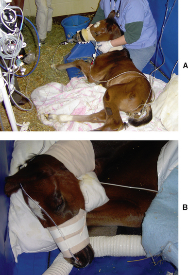
Immediate supportive therapy should begin as soon as the neonate has been transported to the hospital stall. The most important goal of early therapy is assuring tissue oxygen delivery by maximizing pulmonary loading of hemoglobin, guaranteeing sufficient blood oxygen content and returning lung and tissue perfusion to normal. The neonate should be placed on intranasal or mask oxygen insufflation immediately (Figure 7-3, A and B). As the intranasal oxygen line is being placed, but before oxygen is delivered, it is my routine to draw an arterial sample for blood gas analysis while the neonate is still breathing room air. This will be compared to a second sample taken after initial resuscitation. The intranasal oxygen flow is generally begun between 5 and 10 liters/minute to compensate for mismatching if present and also for hypoventilation allowing maximal blood oxygen loading. If the neonate is anemic (PCV <25%), a blood transfusion should be seriously considered. Currently available hemoglobin-based blood substitutes have significant adverse effects in cases such as this and are contraindicated.
Hypoperfusion in the critical neonate is usually secondary to hypovolemia due to poor vascular tone. Sick neonates are almost never dehydrated during the first 48 hours of life unless there is significant diarrhea, reflux, or GI tract pooling of fluids, in rare cases of polyuria, or when there are high insensible losses as in extremely hot weather when tachypnea is present. In fact, when neonates have been subjected to intrauterine stress, because of fluid shifts, they are usually born overhydrated. Critical neonates presenting during the first 48 hours of life are often hyperhydrated but hypovolemic. So when approaching fluid therapy, it is essential to optimize by not only administering high enough volumes to correct the hypovolemia but also to use care not to give excessive fluid volumes that will further exacerbate the hyperhydration.13
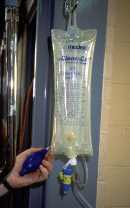
There is no compelling evidence that shows an advantage in using crystalloids or colloids when treating hypovolemia.14 I generally use both with crystalloids playing the dominant role. When choosing a crystalloid, particular attention should be paid to the tonicity and the strong ion difference of the fluid. Since large volumes will be used, there will be a tendency for the plasma osmolarity and strong ion balance to move toward that of the fluids. I prefer isosmotic fluids with a strong ion balance approximating normal plasma. ’04 Revival was given Normosol-R (Abbott Laboratories). Delivering 20 ml/kg boluses of fluids over 10 to 20 minutes allows delivery of fluids in a timely manner but also imposes a set time for reassessment (after completion of each bolus) so as to achieve rapid return of perfusion while avoiding excessive fluid delivery (Figure 7-4). Thus, in a typical 50 kg foal, a 1-liter bolus is given rapidly (usually over 10 minutes with the aid of a pressurized cuff) and once delivered, a rapid assessment of return of perfusion (peripheral body temperature, pulse quality, return of signs of organ function) is used to decide if the bolus should be repeated. One or more liters of plasma are also given for its colloid properties and for the bioactive proteins it contains.
Inopressor Therapy
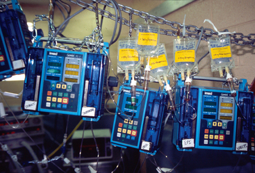
If there is inadequate return of perfusion after four to six fluid boluses or if the patient is deteriorating despite the fluid loading, inopressor therapy should be initiated15 (Figure 7-5). The drugs used most commonly in support of perfusion are adrenergic agonists (usually dopamine, dobutamine, norepinephrine, or epinephrine) and vasopressin, although physiologic doses of corticosteroids, naloxone, and NOS blockers (methylene blue) have also been used. Some of these drugs have more inotropic (increases contractility) effects, while others are more inopressors (increase vascular resistance). When choosing drugs to support the cardiovascular system, it is important to ensure cardiac output. If pressors are used without inotropic support, there is danger that cardiac output and perfusion will decrease (despite an increase in BP numbers). For this reason, inotropes are almost always indicated when pressors are used. Mixed inotropic and pressor support or inopressor support can best be achieved by selecting an inotrope, such as dobutamine or medium-dose dopamine, as part of the initial therapy.16
Stay updated, free articles. Join our Telegram channel

Full access? Get Clinical Tree


