6: THE HINDLIMB

6 THE HINDLIMB


Clinical importance of the hindlimb


The hindlimb can be examined clinically by radiography, by ultrasonography and MRI scanning, and by nerve blocks and anaesthesia of the joints. The major difference between the fore and hind limbs is that in the hindlimb the foot is not the major source of clinical lameness. Also, examination of a hindlimb has many more safety implications than examination of a forelimb. In foals and young horses, trauma and genetics in any combination are the major contributors to lameness problems.


‘Epiphyseal fusion times’ are not always a true indicator of growth plate closures. There are breed variations – generally the growth plates close earlier in the lighter breeds than the heavier breeds. In general, distal growth plates heal earlier than proximal ones. For the femur, the proximal growth plate fuses at 24–36 months and the distal at 24–30 months. For the tibia the corresponding values are 24–30 months and 17–24 months. The lateral malleolus of the tibia has a separate ossification centre, which is shown embryonically to be the distal epiphysis of the fibula. The separate ossification centre for the tibial tuberosity fuses to the epiphysis at 9–12 months of age and to the metaphysis at 30–36 months. The proximal end of the fibula ossifies soon after birth. Two or three separate ossification centres in the fibrous shaft may remain separate for many years. The proximal growth plate of metacarpal bone III is fused at birth and the distal plate fuses at 9–12 months. In foals, the most rapid growth period is from birth to 10 weeks. The activity of the growth plate determines bone length and, to some extent, bone shape. Abnormalities of endochondral ossification may affect bone length and also the axial alignment of the ends of the bones and the shapes of the joints. These effects are visible in the foal.


The hock has six tarsal bones – tibial (talus) and fibular (calcaneus), central, and tarsal bones I, II (fused) and III, IV. The epiphysis of the fibular tarsal bone is evident by 2 weeks and fuses between 24–36 months. Intra-articular fractures of the tarsus are not so common as in the carpus, because the hind limb is not so often involved in trauma.


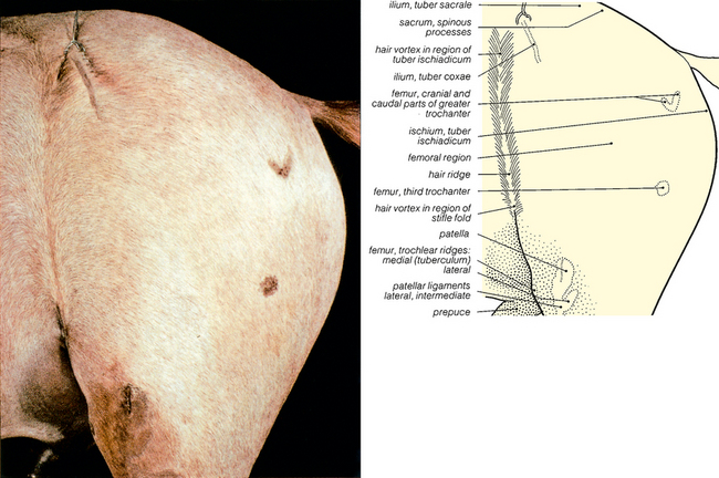
The hip joint is one of the ‘high motion’ joints of the horse, all of these have large synovial spaces which can be entered with a needle. Therefore, anaesthesia of the joint can be used for diagnostic purposes, antibiotics and anti-inflammatory drugs can be administered, and samples can be taken for culture or cytology. The ‘low motion’ joints, for example the tarsus, have small synovial spaces and are bounded by a wealth of collagenous ligaments, tendons and joint capsules. They are therefore much less accessible clinically. Intra-articular analgesia of the coxo-femoral (hip) joint may be used as a diagnostic test. To reach the hip joint, which is not easily palpated because of the massive gluteal muscles, a needle is inserted between the cranial and caudal processes of the greater trochanter of the femur. The needle is directed in a slightly cranial, medial and distal direction to reach the joint. Coxo-femoral luxation does occur, but rarely, and attempts to reduce the dislocation are usually unsuccessful as it is difficult to keep the femoral head in place. Osteoarthritis of the hip joint is rare.


Fractures of the femur are relatively rare and little can be done to repair them. Fracture of the tibia is more common and requires ‘Robert Jones type’ fixation.


The stifle joint is the most common site of pathological problems in the hindlimb. Osteoarthritis is quite common in this joint. The femoral articular cartilage may be 3–4 cm thick and is the thickest in the equine joints. The medial femoral condyle is a common site for bone cysts. Patellar fracture does occur rarely and is usually the result of direct trauma, usually from a kick. Osteochondrosis dissecans is also fairly common in the stifle joint. The stifle joint relies on the particular anatomy of the femur for fixation in extension. The medial ridge of the femoral trochlea can act as a ‘catch’ for the patellar fibrocartilage and the medial patellar ligament, enabling the joint to be locked in extension. One of the most common injuries of the stifle is upward fixation of the patella, diagnosed by palpation and radiography. In this condition the patella does not spontaneously unlock when attempts are made to flex the joint. Problems may also occur when the lateral femoral trochlear ridge is hypoplastic and there is no ridge to hook over the patellar ligaments. It is often congenital and in that case it is often bilateral. The femoro-patellar joint communicates with the medial femoro-tibial joint and, in 25% of horses, also with the lateral femoro-tibial joint. In the other 75% of horses the lateral femoro-tibial joint is separate. The two synovial cavities of the femoro-tibial joint do not directly communicate with each other. The seat of lameness can be detected by anaesthesia of these synovial cavities. The femoro-patellar joint is approached for intra-articular analgesia by inserting a needle proximal to the tibial crest, lateral or medial to the middle patellar ligament, with the needle directed slightly proximal. The medial femoro-tibial joint is approached proximal to the tibia, with the needle inserted between the medial patellar ligament and the medial collateral ligaments. The lateral femoro-tibial joint is approached proximal to the tibia, with the needle inserted caudal to the long digital extensor tendon and cranial to the lateral collateral ligament. The femoro-tibial joint is the site of a wide variety of stifle injuries including those to the menisci, the articular cartilage, the collateral ligaments and the cruciate ligaments (commonly caused by twisting of the stifle). All of these can be investigated by arthroscopy under general anaesthesia.


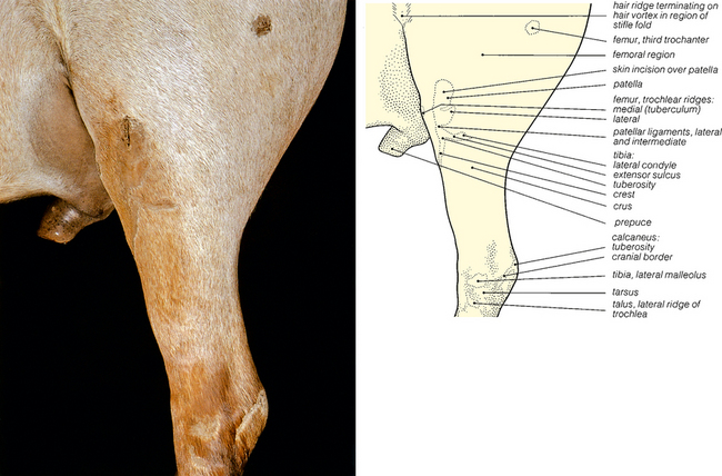
There are four synovial sacs in the hock. The first is in the tarso-crural joint. The second is in the proximal intertarsal joints (this communicates with the sac of the first joint). The third is in the distal intertarsal joints (this communicates with the sac in the tarso-metatarsal joint) and finally there is the synovial sac of the tarso-metatarsal joint. The hock can be investigated using intra-articular analgesia. The proximal joint (tarso-crural) is entered by inserting a needle either side of the saphenous vein, just distal to the medial malleolus. The distal intertarsal joint can be found on the medial aspect of the hock, just distal to the proximal border of the medial branch (cunean) of the tendon of the cranial tibial muscle. The needle is inserted in a lateral direction, horizontally, between the third and central tarsal bones. The tarso-metatarsal joint is approached via a small depression proximal to the head of the lateral splint bone (metatarsal bone IV). A needle is inserted in a horizontal and slightly downward direction to a depth of approximately 2–3 cm.


Osteoarthritis of the small hock joints does occur. ‘Bog spavin’ is a descriptive term for synovial effusion of the tarso-crural joint. It is the result of a low-grade synovitis. The swelling appears in the proximo-medial part of the hock. It is not a major cause of persistent hindlimb lameness in the horse. Osteochondrosis dissecans, a degenerative condition of cartilage, also affects the hock, particularly the distal intermediate ridge of the tibia and the lateral trochlear ridges of the tibio-tarsal joint. The hock joint can also suffer from bacterial infection and inflammation, similar to gonitis of the stifle joint. This often results in extensive necrosis and ultimately joint collapse. Another important condition of the hock is fluid distension of the tarso-crural joint which occurs dorso-medially between the fibularis (peroneus) tertius muscle and the medial malleolus; dorso-medially between the medial malleolus and the deep digital flexor tendon; and caudo-laterally between the lateral malleolus and the tuber calcis. Tarso-crural dislocation (luxation) is a rare event which is virtually impossible to repair and is an indication for immediate euthanasia.

A considerable amount of clinical time is spent dealing with the problems associated with the muscles and tendons of the hindlimb. Rupture of the fibularis (peroneus) tertius muscle (which runs from the extensor fossa of the distal femur to the dorso-lateral aspect of metatarsal bone III, the calcaneus and tarsal bones III, IV) occurs when there is overextension of the hock joint, usually in racing. It is important, as this tendinous muscle is part of the hindlimb stay apparatus, which enables the hock and stifle to flex or extend in unison. ‘Capped hock’ is distension of the subcutaneous bursa overlying the superficial digital flexor tendon (SDFT) at the point of the hock. It is usually only of cosmetic significance. It results from trauma. ‘Deep capped hock’ is damage to the large synovial bursa underneath the tendon, resulting in swelling on both sides of the tendon. Traumatic injury to the semitendinosus muscle results in scarring with adhesions between the semitendinosus and semimembranosus muscles. Rupture of the gastrocnemius muscle can occur and tendinitis of the muscle is also a possibility. The muscles of the hindlimb that are injured most often are the major flexor muscles together with their tendon sheaths and suspensory ligaments. The SDFT is easily damaged and may be partially dislocated (sub-luxated) when the horse is in work. Most commonly, this occurs laterally but sometimes medially. The problem is made much worse when the tendon is also split. Surgical correction of this is generally unrewarding. There is also a specific condition known by the colloquial name of ‘curb’ which is desmitis (inflammation) of the SDFT. It is seen clinically as a swelling on the plantar aspect of the hock, approximately 10 cm distal to the point of the hock. ‘False curb’ is a swelling or enlargement on the proximal aspect of the splint bones (metatarsal bones II and IV). The extensor muscles and their tendon sheaths can also be damaged, but in these cases the injuries are not so severe, and conservative treatment is usually successful. Injuries to the suspensory apparatus also occur. Lesions confined to the proximal third of the metatarsus are usually referred to as proximal suspensory desmitis. Here there is localized oedema, swelling, heat, distension of the medial palmar vein and possibly pain. The condition may be mild, moderate or severe and can become chronic. More rarely there are avulsion (‘tearing’) fractures of the whole tendon and complete breakdown of the tendon. Sub-tarsal analgesia and ultrasonography can be used for diagnosis.


Fluid distension of the tarsal sheath of the deep flexor tendon is known as ‘thoroughpin’. It is usually caused by trauma. The sheath begins approximately 5–7 cm proximal to the level of the medial malleolus and extends distally to the upper third of the metatarsus. Swellings can be found on either side of the common calcaneal tendon and cranial to the tendon.


The femoral nerve can be damaged by penetrating wounds of the caudal flank which result in abscesses. The nerve is also damaged rarely by tumours, aneurysms, and pressure during parturition. Since it innervates the quadriceps group, which flexes the hip and extends the stifle, forward movement is limited. The cranial gluteal nerve may be damaged by trauma; if this happens atrophy of the gluteal muscles may follow. The sciatic nerve can be damaged by systemic bacterial infections such as salmonellosis, but also by infectious processes extending from the sacrum and the pelvis. Damage will affect both tibial and fibular (peroneal) nerves.


Paralysis of the tibial nerve, which innervates the extensors of the tarsus and the flexors of the digits and stifle, is not uncommon. Diagnostic blocking can be achieved by injection 10 cm proximal to the tuber calcis on the caudo-medial aspect of the limb, in the space between the deep digital flexor tendon and the common calcaneal tendon. The nerve lies beneath the superficial fascia, about 1 cm deep, and the needle should be directed just cranial to the common calcaneal tendon, with care not to penetrate the skin on the lateral aspect.


The fibular (peroneal) nerve innervates the flexors of the tarsus and the extensors of the digit. It can be blocked 10 cm proximal to the point of the hock, in a groove between the long and lateral digital extensor muscles, just above the musculo-tendinous junction. This is proximal to the lateral malleolus of the tibia, on the lateral aspect of the thigh. The deep branch of the fibular (peroneal) nerve can be blocked 2–3 cm below the superficial fascia and the superficial branch 1–2 cm below the fascia.


Below the hock, the plantar nerves can be blocked between the deep digital flexor tendon and the suspensory ligament. The plantar metatarsal nerves can be ring-blocked on the proximal metatarsus. The plantar metatarsal nerves are blocked axial to the splint bones, abaxial to the suspensory ligament, and along the plantar cortex of metatarsal bone III.


image

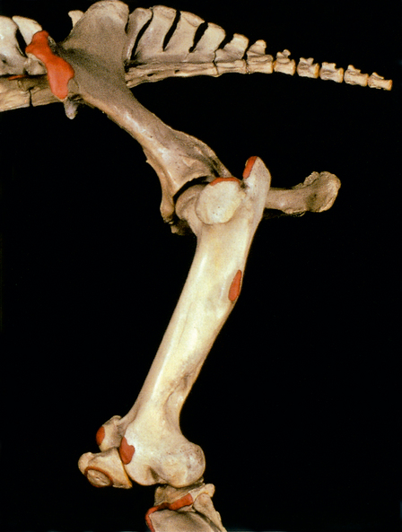
Fig. 6.2 Bones of the pelvic and femoral regions: left lateral view. The palpable bony features shown in Fig. 6.1 have been coloured red, except the tuber sacrale and the sacral spines. In this skeleton the patella has not been positioned so distally in the trochlea: in the embalmed horse the hindlimb was fully extended before nailing the patella to the trochlea.


< div class='tao-gold-member'>
Stay updated, free articles. Join our Telegram channel

Jul 8, 2016 | Posted by in SUGERY, ORTHOPEDICS & ANESTHESIA | Comments Off on 6: THE HINDLIMB

Full access? Get Clinical Tree

Get Clinical Tree app for offline access