Fig. 1.
Ten-microlitre microsyringe setup for MFB lesion injections. The 30-gauge cannula is connected to the needle of the syringe using polythene tubing. The syringe, tubing and cannula are filled with sterile infusion saline prior to use (priming).
3.2 Detailed Surgical Methodology
3.2.1 Unilateral Lesion of the MFB
Table 1 gives two sets of coordinates; the first is based on Dunnett et al. (9) and is designed for use on rats over 250 g in weight. The second is based on recent modifications made by Torres et al. [38] and is recommended for rats in the 150–250 g weight range.
Table 1
Stereotaxic coordinates for MFB injection of 6-OHDA
Coordinates from bregma (mm) | ||||
|---|---|---|---|---|
Nose bar | A= | L= | V=a | |
MFB lesion I | −2.3 | −4.4 | −1.0 | −7.8 |
MFB lesion II | −4.5 | −4.0 | −1.2 | −7.0 |
1.
Anaesthetize the rat, and place the rat in the stereotaxic frame with the nose bar set at the appropriate height (see Table 1).
2.
Shave the scalp using hair clippers, and make an incision using a scalpel, through the skin along the midline (maximum 2.5 cm long) starting from just behind the inter-ocular line, and cutting caudally.
3.
Open the incision and use the scalpel blade to pare back the underlying connective tissue and scrape the exposed surface of the skull clean.
4.
Position the tip of the drill bit directly over bregma; the zero point for x – y coordinates.
5.
Raise the drill bit from the skull and move it to the x – y coordinates (see Table 1). Drill through the skull, taking care not to damage the underlying dura matter.
6.
Load the cannula with 4–5 μl of 6-OHDA, first drawing a small “spacing” air bubble (0.5–1 μl) into the cannula to prevent mixing of the neurotoxin and the priming solution.
7.
Lower the cannula into the drilled hole until it is just touching the dura mater. If the dura matter is undamaged, the correct depth is determined when the meniscus of fluid in the drill hole descends slightly, as the dura membrane is touched by the cannula.
8.
Lower the cannula into the brain, allowing the cannula to pierce the dura mater to the desired depth from the dura mater.
9.
Start the syringe pump and run for 3 min to deliver 3 μl of neurotoxin. Smooth flow of the toxin without blockage of the cannula is monitored by movement of the spacing bubble in the transparent polyethylene tubing.
10.
Stop the pump and leave the cannula in place for a further 2 min to allow fluid diffusion, before slowly retracting from the brain.
11.
Purge any unused neurotoxin from the cannula and flush several times by repeated drawing and expulsion of clean saline solution.
12.
Suture the skin and administer analgesia according to the local SOP.
13.
Place animal in a heated recovery cage until fully conscious. With gaseous anaesthesia this usually takes 5–10 min, after which the rat can be returned to its home cage.
The 6-OHDA has an immediate effect; recovering animals will have a postural bias towards the lesion side and rotate ipsilaterally. Checking for rotation in recovering animals is the first index of lesion success. Post surgery complications with this type of lesion are very rare but animals should be given a thorough check 24 and 48 h later.
3.2.2 Partial Lesions by Striatal Injection of 6-OHDA
Table 2 shows coordinates for multiple injection of the toxin into the striatum. The coordinates are based on those described by Kirik et al. (4) modified so that the four injection sites are in a straight line, 1 mm apart, enabling four cannulae (each attached to a different syringe) to be used simultaneously. This requires a cannula holder like the one shown in Fig. 2 that has vertical grooves, exactly 1 mm apart, in which the cannulae are held tightly at the correct spacing. The cannulae are adjusted to the same length in the cannula holder by loosening the clamp and lowering the cannula tips onto the top surface of the ear bar before re-clamping. If a single syringe and cannula are used, drill all four holes before making the first injection. Note that, because the drill holes are evenly spaced, after positioning and drilling the first hole, movement between subsequent drill holes is always the same: A = −0.6, L = −0.8.
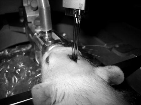
Table 2
Stereotaxic coordinates for striatal injection of 6-OHDA
Coordinates from bregma (mm)a | |||
|---|---|---|---|
A= | L= | V= | |
Rat Striatal lesion b | +1.0 | −2.0 | −5.5, −5.0, −4.5 |
+0.4 | −2.8 | −5.5, −5.0, −4.5 | |
−0.2 | −3.6 | −5.5, −5.0, −4.5 | |
−0.8 | −4.4 | −5.5, −5.0, −4.5 | |
Fig. 2.
Four-cannula setup for simultaneous injection of 6-OHDA at four sites in the striatum.
2.
Shave the scalp and make an incision in the skin along the midline (maximum 2.5 cm).
3.
Open the incision and clean the surface of the skull with a scalpel blade.
4.
Position the tip of the drill bit over bregma.
5.
Move the drill bit to the x – y coordinates for the first hole (see Table 2) and drill through the skull, taking care not to damage the underlying dura matter.
6.
Repeat the process for holes 2–4.
7.
Load each cannula with 3–4 μl of 6-OHDA, leaving a small air bubble in each syringe setup, between the neurotoxin and the priming solution.
8.
Lower the cannulae until just touching the dura matter (see above).
9.
Lower the cannula into the brain to the maximum depth from the dura matter.
10.
Start the syringe pump and run for 2 min, raising the cannula(e) 0.5 mm after 40 s and again after 1 m 20 s to deliver 2 μl of neurotoxin at three different levels per injection.
11.
Stop the pump and leave the cannula(e) in place for a further 2 min, before slowly retracting from the brain.
12.
Flush the cannulae as above.
13.
Suture the skin and administer analgesia according to your local SOP.
14.
Place animals in a heated recovery cage until fully conscious.
3.3 Rotation Testing
In our laboratory, operated rats are tested for lesion efficacy using amphetamine (methamphetamine hydrochloride), at 2 weeks and 4 weeks post-lesion, and using apomorphine (apomorphine hydrochloride dehydrate) at 5 weeks post-lesion. Rotation is usually assessed using an automated rotometer system, over a 90 min test session for amphetamine and a 60 min session for apomorphine. Immediately following injection, rats are placed in 30 cm diameter circular, flat bottomed bowls, enclosed in 30–50 cm high Perspex or aluminium cylinders. Rats are attached via a harness to a rotometer head which sends information to a computer-controlled, automated rotometer system (e.g. Rotomax System, AccuScan Instruments Inc.). The system records both ipsilateral and contralateral turns, from which the net rotation (ipsilateral minus contralateral) can be calculated. Rota-tion scores are reported as either net rotations over the entire session, or as mean net rotations per minute. When an automated system is not available, alternative methods of measuring rotation may be used. Method one is to make a video recording of each animal’s rotation session and then count rotations from the recording using high speed playback. A second method is to sample the rotation of each animal by recording rotation at regular intervals over the session. Animals are observed for 1 min at a time at either 10 min or 15 min intervals, recording both ipsilateral and contralateral rotations. Scores may then be extrapolated for the entire session.
In our early studies, we used a minimum criterion for lesion success as seven turns per minute under 5 mg/kg methamphetamine. However, this dose was associated with high levels of stereotypic behaviour and occasional mortality, As a result, we now use a somewhat lower dose (2.5 mg/kg), and six net rotations per minute (540 net rotations in a 90 min session) as the criterion for a successful 6-OHDA lesion corresponding to approximately 95% depletion of striatal dopamine in studies based on post-mortem neurochemical assay.
Re-lesioning of rats which do not meet this criterion is seldom successful, and as a result they are usually excluded from the experiment. For subsequent treatments, lesioned rats are allocated into counterbalance groups matched either on the 4-week amphetamine, or the 5-week apomorphine scores, such that prior to treatment, all groups have approximately the same mean rotation scores.
Following experimental treatments to ameliorate or reverse the effects of the 6-OHDA lesion, rotation scores are also used for in vivo assessment of treatment efficacy. Pre-lesion, protective treatments, such as growth factors (e.g. GDNF) or anti-apoptotic agents (e.g. caspase inhibitors), may reduce lesion-induced dopamine cell death in the ventral midbrain (10, 11). This is reflected in reduced levels of amphetamine rotation in the treated groups. Post-lesion treatments such as replacement of dopamine function by implantation of embryonic dopamine cells, or dopaminergic cells derived from stem cells, restore dopamine innervation to the lesioned striatum, and reduce the levels of net ipsilateral rotation. Interestingly, large dopamine grafts in the striatum can cause a net contralateral rotation (so-called over-compensatory rotation) due to their action on residual supersensitive receptors in the ipsilateral striatum (12, 13).
3.4 Behavioural Assessment
Unilateral lesions of the dopamine system induce well characterized deficits on the contralateral side of the body. The principal methods used to assess these deficits include the stepping, cylinder, staircase, and corridor tests, all of which detect either preferred use of the ipsilateral limbs or neglect of contralateral space. For full details of behavioural testing of unilateral lesioned animals, see the chapters by Fleming and Schallert, Smith, and Heuer, this volume and Farr and Trueman Volume II in this series.
3.5 Post-Mortem Assessment
The efficacy of the 6-OHDA lesion may assess post-mortem using immunohistochemical detection of the dopamine synthesizing enzyme, tyrosine hydroxylase (TH). Successful MFB lesions induce massive loss of TH immunoreactive neurons in the ipsilateral substantia nigra compacta and ventral tegmental area. Following striatal injection of the toxin, the majority of SNc neurons are lost. Fixation, sectioning, and staining of brain tissues are beyond the scope of the current chapter. Briefly, animals are sacrificed and perfused transcardially using formaldehyde fixative (usually 4% in phosphate-buffered saline). Frozen sections may then be stained immunohistochemically using antibodies against tyrosine hydroxylase. For a detailed methodology, see Torres et al. (14).
< div class='tao-gold-member'>
Only gold members can continue reading. Log In or Register to continue
Stay updated, free articles. Join our Telegram channel

Full access? Get Clinical Tree


