4: THE THORAX

4 THE THORAX


Clinical importance of the thorax


The thorax contains two major organs that are of considerable importance in clinical medicine in the horse – the lungs and the heart – but the nostril is the point at which physical abnormalities of the respiratory tract such as blood, excess mucus, or a purulent nasal discharge, are seen. The real test in clinical diagnosis is to ascertain whether this abnormality is coming from the upper or lower respiratory tract and from which component part of either.


Other physical abnormalities of respiration are most commonly detected by watching (abnormal excursions of the thoracic walls) or listening (coughing, roaring or rales) to the breathing and it is usually these that will necessitate further, more detailed, examination of the respiratory tract. One of the most common findings is dyspnoea (difficult breathing) which is seen in ‘heaves’ (pulmonary emphysema); typically, the affected horse has a second expiratory effort. The respiratory tract includes a variety of clinically important components such as the nasal cavity, nasopharynx, larynx, and guttural pouches, as well as trachea and lungs. Examination of the mucous membranes of the eye and the mouth (paleness, blue discolouration, abnormal capillary refill time) may suggest general abnormalities of the cardiovascular system such as anaemia or hypoxia.


The arterial pulse will give information on the heart and circulation. This can be taken from several arteries, including the transverse facial, facial, digital, brachial, femoral and the external carotid. Also, the aorta can be palpated per rectum. In the horse there is also a jugular pulse, which can be seen from the thoracic inlet to halfway up the neck when the horse is in the normal standing position.


The area of the chest available for clinical examination is limited by the presence of the forelimb, thoracic musculature and the decreasing thickness of the lung in its caudal parts within the chest. The effective area for listening is bounded caudally by a line that runs from the 18th rib to the middle of the thorax at rib 13, to shoulder level at rib 11, and finishes at the point of the elbow. For both organ systems, further evaluation is carried out using auscultation, percussion, MRI scanning (upper respiratory tract and sinuses), endoscopy, radiography (mainly in foals due to size limitations and X-ray power required in adults) and laboratory-based evaluations. A wide variety of diagnostic techniques can be applied to the thorax. Lung biopsy is not widely used; less invasive techniques have replaced it and pneumothorax sometimes followed its use. If it is necessary to perform lung biopsy, the ventral half of intercostal spaces 7–10 can be used. Endoscopy is widely used for the collection of samples from all levels of the respiratory tract. Tracheal and broncho-alveolar lavage (BAL) can be used to obtain samples for microbiology and cytology from as low as the 4–5th generations of the bronchial tree, using sterile saline. Tracheal aspirates can be drawn directly from the trachea by inserting a sterile needle between the tracheal cartilages. Thoracocentesis (removal of fluid from the chest) uses intercostal spaces 6–9 on either side of the chest, with spaces 7–8 being the best. The site is just above the costo-chondral junction. The caudal part of the intercostal space is used, as the intercostal artery, vein, lymphatics and nerve lie in the rostral part. During this procedure, care is needed to avoid the superficial thoracic vein. Pleural fluid can be collected for cytology and bacteriology.


There are many examples of infectious respiratory disease including, in former times, the bacterial zoonosis, glanders. Strangles, associated with streptococcal infection (S.equi subspecies equi) is one of the most serious disorders, localising in the lymph nodes of the respiratory tract and causing purulent discharges and abscesses. False strangles is similar and is caused by S. zooepidemicus. Other bacterial infections of the respiratory tract include pneumonia associated with Rhodococcus equi, particularly in foals and young horses where there may be up to an 80% mortality. This disease can also lead to a severe arthritis with antigen/antibody complexes in the joints of young horses. A second category of pulmonary disease is associated with parasites including parasitic pneumonia caused by the migration of P.equorum and also lungworms such as Dictyocaulus arnfeldii (commonly picked up from donkeys, which are carriers of the infestation) and, occasionally, pulmonary hydatidosis. There are several severe viral infections of the equine respiratory tract. One of the most important is equine influenza. Equine herpes virus may cause upper and lower tract disease, specific laryngitis, pharyngitis and abortion. Other important viral diseases include rhinovirus infections, equine viral arteritis, African horse sickness and equine infectious anaemia (swamp fever). Pulmonary oedema, acute respiratory distress syndrome and aspiration pneumonia are also not unknown.


Non-infectious respiratory disease is also important. Epistaxis (nosebleed) occurs in approximately 5% of racehorses after galloping. Exercise-induced pulmonary haemorrhage occurs after exercise. Reversible airway obstruction occurs with an asthma-type equivalent which can also be found on summer pastures. Chronic obstructive pulmonary diseases (now called RAO – recurrent airway obstruction, COPD, ‘heaves’) are caused by allergy (hypersensitivity) to dust allergens. Occasionally the horse may suffer from amyloidosis, pulmonary effusions (these can be checked by thoracocentesis) and pneumothorax, which can be alleviated by inserting a cathether high in the 13th intercostal space and aspirating the air. As well as abnormalities of the heart, the equine thorax may also suffer damage to great vessels as in equine viral arteritis, thrombophlebitis, or purpura haemorrhagica. Arterial rupture and aneurysm (particularly of the internal carotid artery) have been known to follow damage to the arteries from migrating nematode larvae.


Equine cardiology is an important subject, with auscultation as the most important clinical technique. In addition, an electrocardiogram (ECG) or 12-lead ECG can be used for the diagnosis and assessment of the severity of valvular, pericardial, myocardial and great vessel disease. Up to 4 heart sounds may be heard in normal horses. The first, which marks systole, is the closure of the atrioventricular (A-V) valves and opening of the semi-lunar aortic and pulmonary valves. The left atrioventricular valves are best heard in the left 5th intercostal space, midway between the level of the point of the shoulder and the point of the elbow. The right A–V valves are best heard in the right 4th intercostal space, midway between the level of the point of the shoulder and the point of the elbow. The second sound, which marks diastole, is caused by the closure of the semilunar valves and the opening of the atrioventricular valves. The aortic valves can be heard in the left 4th intercostal space at the level of the point of the shoulder. The pulmonary valves are heard on the right side in the 4th intercostal space, midway between the level of the point of the shoulder and the point of the elbow. The third heart sound occurs early in diastole and is heard in only one-third of horses. It marks the point at which the blood decelerates, at the end of rapid ventricular filling. The fourth sound, audible in most horses, is the atrial contraction in late diastole.


There are various cardiac complaints in the horse. Congestive heart failure can be left-sided or right-sided. Pericarditis, pericardial effusions, and myocarditis (ionophore toxicity) occur. Aortic root rupture results in sudden death with massive haemorrhage into the thoracic cavity. Many abnormal sounds may be heard in the examination of the heart. They include cardiac murmurs which can indicate stenosis of valves, incompetence of valves, shunts, and pre-systolic and systolic murmurs. Ectopic beats also occur in some horses. Disorders of cardiac rhythms include sinus arrhythmia, sinus tachycardia, ventricular tachycardia, atrial fibrillation, ventricular fibrillation, atrio-ventricular block and sino-atrial block. Congenital heart defects do occur rarely in foals. These include ventral septal defects, persistent ductus arteriosus, patent foramen ovale, myocarditis and fibrocarditis.


Clinical considerations for the spine in the thoracic region are dealt with in the section on the spine in Chapter 8 (p. 269).


image

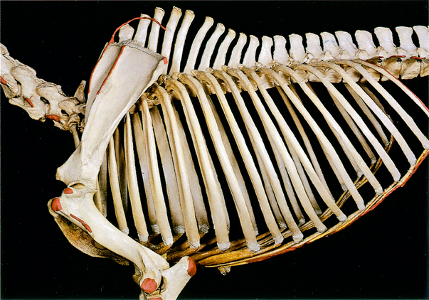
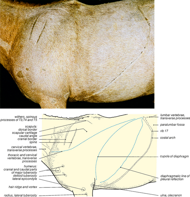
Fig. 4.2 Skeleton of proximal forelimb and thorax: left lateral view. The palpable features shown in Fig. 4.1 are coloured red except for the spinous processes at the withers. Note that in this articulated skeleton rib 18 is large. In the dissected specimen this rib was small (see Fig. 4.9) and the last distinctly palpable rib was rib 17.


< div class='tao-gold-member'>

Stay updated, free articles. Join our Telegram channel

Jul 8, 2016 | Posted by in SUGERY, ORTHOPEDICS & ANESTHESIA | Comments Off on 4: THE THORAX

Full access? Get Clinical Tree

Get Clinical Tree app for offline access