3: THE FORELIMB

3 THE FORELIMB


Clinical importance of the forelimb


The foal grows rapidly, especially from birth to 10 weeks-of-age. Fusion of growth plates in the limb bones gives an approximate estimation of the age of the young horse. Lighter breeds tend to fuse earlier than the heavier breeds. The proximal humeral growth plate closes at about 24–42 months of age and the distal at 12–24 months. In the radius, the proximal growth plate fuses at 12–24 months of age and the distal plate at 24 months (it has continuous growth for the first 60 weeks of life). The ulnar growth plates fuse at around 24–36 months. The proximal growth plate of metacarpal bone III is fused at birth, as are the distal growth plates of phalanges I and II. The distal growth plate of metacarpal bone III fuses at 6–9 months and the proximal growth plates of phalanges I and II fuse around 6–12 months.


In the young horse there may be abnormalities of conformation and if there are angular bone abnormalities they are best sorted out by 70 days of age. They are usually crooked legs, with flexure and contractile deformities. The young horse may also suffer from osteochondrosis dissecans in the shoulder and elbow and this may be unilateral or bilateral. Sub-chondral bone cysts may also affect the young horse.


Equine limbs are often subjected to trauma, especially the forelimbs.


It is not easy to separate the components that may be involved in the complex subject of lameness. It is important to localise the site(s) of the problem and to characterise the nature of the pathological changes. This requires a good history, an overall inspection, a complete physical examination which includes palpation and manipulation and, ultimately, a detailed evaluation. Clinical examination will reveal deformity, swellings or thickenings, skin wounds, and muscle wasting.


Palpation detects heat and pain as well as the precise location and consistency of swelling. Superficial pain is usually tested by poking the skin and deep pain is tested by using hoof testers, limb flexion or deep digital palpation. Analgesia is used for joints and also for diagnostic nerve blocks. In chronic disorders, the induced analgesia may not completely block out the diagnostic lesions. If more than one site, or both limbs, are involved it may be even more difficult. After using analgesia on the nerves in the upper parts of the limbs, loss of motor function and stumbling may result. Manipulation of the joints allows evaluation of movement and detects restrictions, instabilities, pain and crepitus. Abnormalities of gait may be detected. These results can then be further investigated by the available techniques such as radiology, ultrasonography, scintography, magnetic resonance imaging, computed tomography and the use of regional anaesthesia and specific nerve blocks to localise the site of lameness.


There are several types of fractures. The direction and location of the fracture line within a single bone may have a significant effect on the feasibility of repair. The relative displacement of the fractured pieces is also important. In some instances the fracture is incomplete. It may be stable or unstable, but probably the most important feature is whether it is open or closed, as an open fracture is almost invariably infected. The treatments of individual fractures are beyond our remit but fractures of proximal limb bones are difficult, often impossible, to repair and distal limb fractures are more accessible. In nearly all fracture cases there is an acute, non-weight-bearing lameness. Techniques of diagnosis and treatment continually improve, but the fact that they all require formation of a callus, followed by subsequent remodelling, will never change. Anaesthetic recovery and aftercare for these fractures following surgical repair, are the most crucial aspect of equine orthopaedics.


The scapula (four centres of ossification) is not easily fractured, but the tuber scapulae can be fractured and the articular surface of the glenoid cavity may be affected. The proximal humerus has three centres of ossification which fuse together at about 3–5 months of age; the distal humerus has two centres. This bone also is not commonly fractured as, like the scapula, it is supported by strong musculature, but all the other bones of the forelimb are more exposed.


A fractured radius requires a Robert Jones bandage to prevent adduction. Fracture of the ulna often involves the articular surface of the trochlear notch. This, and separation of the olecranon (also not uncommon following a fall or being kicked by another horse) are both seen as ‘dropped elbow’ because the triceps muscle is unable to prevent it.


Fracture of the accessory carpal bone and chip fractures of the carpus may lead to carpal sheath injuries.


The activities of the horse predispose to a wide variety of joint lesions, often associated with the extra stresses which result from galloping and jumping. There are a variety of joint diseases, including idiopathic synovitis, traumatic arthritis, and osteoarthritis. Osteoarthritis may be primary or secondary following mechanical disruptions and cytokine-mediated inflammation (antibody-associated, free radical damage, or enzyme damage). The initial sign is nearly always a result of extra fluid accumulation in the joint and appears as a soft tissue swelling around the joint. Most of the joints with great mobility have a large joint capsule from which fluid is easily aspirated, but ‘low motion’ joints, such as those of the carpus, are much more difficult to aspirate.


These synovial structures may also be affected by external puncture wounds. Quite often there is a small wound, followed 2–3 days later by swelling, followed by synovial fluid leaking from the joint. Joints are also affected by haematogenous spread of organisms (particularly in foals), local penetration, or local extension from peri-articular structures.


Arthrocentesis followed by intra-articular medication is commonly performed when diagnosing and treating joint disease. Synovial fluid analysis aids in the interpretation of joint disease and is particularly important in horses with septic arthritis. Synovial fluid analysis requires visual assessment, examinations of volume, total protein, inflammatory cells and a cytological analysis.


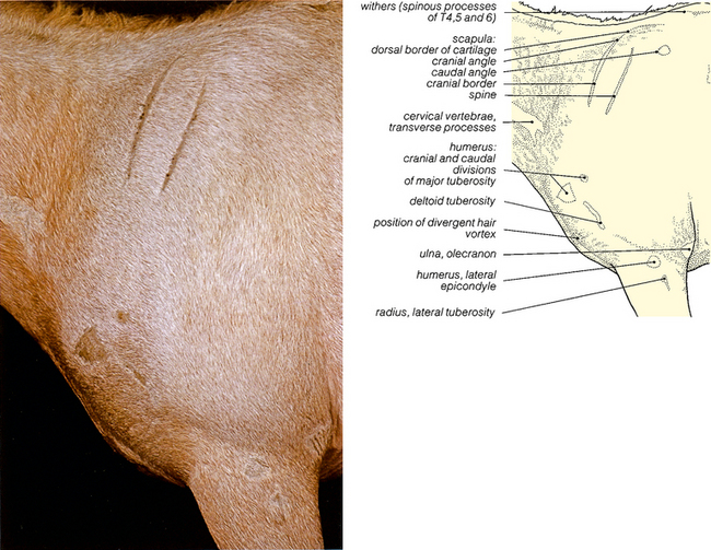
The shoulder joint may show luxation or sub-luxation, particularly if there is laxness in the ligaments of the joint and tendons of the muscles. Intra-articular analgesia of the shoulder joint requires access between the cranial and caudal prominences of the greater tubercle of the humerus, with the needle directed in a horizontal and slightly caudo-medial direction, to enter the synovial cavity.


The elbow joint is very large and is often affected by septic arthritis. Capped elbow is an acquired bursa at the point of the elbow. Intra-articular analgesia of the elbow joint is possible. The lateral humeral epicondyle and the lateral tuberosity of the radius are palpated, and the needle inserted between them, cranial or caudal to the lateral ligament, in a horizontal direction, to a depth of 4–5 cm.


Carpal joint disease is quite common. There are 7 or 8 bones in the carpus. The proximal carpal row has radial, intermediate, ulnar and accessory carpal bones. The distal row has carpal bones II, III, IV and sometimes carpal bone I. There are approximately 26–27 separate articulations between these bones. There are essentially three joints – the antebrachio-carpal, which both rotates and glides; the middle carpal joint which is a simple hinge joint and the carpo-metacarpal articulation which has very little movement.

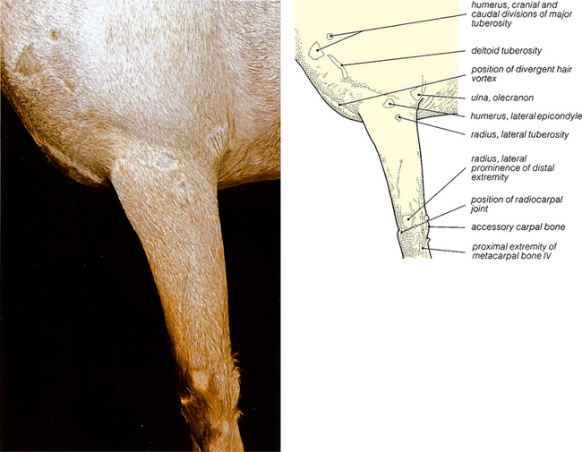
Intra-articular analgesia of the carpal joints is possible as a diagnostic technique. With the limb fixed in a flexed position, the injection sites are easily palpable. The radio-carpal joint is found by locating the depression between the radius and the proximal row of the carpal bones. For the middle carpal joint, locate the depression between the proximal and distal rows of the carpal bones. For both joints the needle is inserted medial to the extensor carpi radialis tendon or between that tendon and the common digital extensor tendon.


Muscles and tendons, and the synovial structures associated with them, are very commonly subjected to injury in any type of working horse. Rupture of the common digital extensor, usually in the sheath over the lateral aspect of the carpus, can occur in foals with carpal contracture. The fibrous palmar carpal joint capsule is dense, and closely attached to the palmar aspect of the carpal bones. It forms the dorsal wall of the carpal canal. It continues distally to form the accessory head of the deep digital flexor tendon (the accessory or inferior check ligament). The carpal fascia on the palmar aspect of the carpal region forms the flexor retinaculum (transverse carpal ligament) between the free palmar edge of the accessory carpal bone and the medial aspect of the carpus. This completes the carpal arch, which contains the superficial digital flexor and deep digital flexor tendons. Carpal flexor tendon sheath injuries are less common than injuries to the digital flexor tendon sheath, but can be a severe cause of lameness. They often result from fractures of accessory carpal bones. The tendon sheath extends from 4–8 cm proximal to the accessory carpal bone to 5–10 cm distal to the carpal bone. The proximal sheath is caudal to the distal radius and cranial to the deep digital flexor tendon. The accessory ligament of the superficial digital flexor tendon (radial or superior check ligament) forms part of the medial wall of the sheath. The short radial head of the deep digital flexor muscle protrudes into the sheath proximally and is inserted into the dorso-medial aspect of the deep digital flexor tendon, approximately 2 cm proximal to the accessory carpal bone. Midway between the proximal recess of the sheath and the accessory carpal bone, caudal to the distal radius, the sheath extends laterally and medially to the deep digital flexor tendon.


Damage to the nerves of the forelimb is usually caused by trauma from normal activities or by aggression from other horses. Damage to the suprascapular nerve, which is motor to the supraspinatus and infraspinatus muscles, results in ‘shoulder slip’. Atrophy of the shoulder muscles may take 18 months to recover.


Radial nerve damage may accompany humeral fractures. The loss of innervation to the extensor tendons of the digit causes a dropped elbow and inability to stand, resulting in long periods of lateral recumbency. Damage to the ulnar and median nerve may result in the loss of skin sensation to palmar metacarpal and to palmar and dorsodistal digital regions. Analgesia of the antebrachium can be carried out by blocking the median nerve medially, 5 cm distal to the elbow joint, where the nerve runs along the caudal aspect of the radius. The ulnar nerve is blocked 10 cm proximal to the accessory carpal bone, in a groove between the flexor carpi ulnaris and the extensor carpi radialis (ulnaris lateralis) muscles.


Damage to the musculocutaneous nerve results in an inability to flex the elbow, as there is no effective innervation of the biceps brachii, coracobrachialis and brachialis muscles. There is also a loss of sensation to the dorsomedial aspect of the limb from the carpus to the fetlock. The nerve can be blocked subcutaneously on either side of the cephalic vein, about half way between the carpus and the elbow, to anaesthetise both cranial and caudal branches of the metacarpal nerves.


The axillary nerve may be damaged. This results in reduced flexion of the shoulder and lateral instability of the shoulder joint, as there is no effective innervation of deltoid and teres major and minor muscles.


image

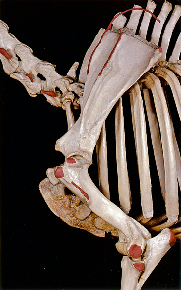
Fig. 3.2 The bones of the forelimb. Scapular and brachial regions: left lateral view. The palpable bony prominences shown in Fig. 3.1 have been coloured red. The palpable edge of the scapular cartilage is shown by a red wire. The tips of the spinous processes at the withers have not been coloured.


< div class='tao-gold-member'>

Only gold members can continue reading. Log In or Register to continue

Stay updated, free articles. Join our Telegram channel

Jul 8, 2016 | Posted by in SUGERY, ORTHOPEDICS & ANESTHESIA | Comments Off on 3: THE FORELIMB

Full access? Get Clinical Tree

Get Clinical Tree app for offline access