13 Cardiac Disorders
Case 13-1 Foal with Congenital Cardiac Defect
’01 Dear John, a 21-day-old Arabian filly presented for evaluation of a loud heart murmur audible on both sides of the chest, and a palpable precordial thrill. ’01 Dear John’s birth 21 days prior to admission to a maiden mare was uncomplicated, and the foal stood and nursed within two hours of birth. Her IgG level was >800 mg/dl. The foal was active and similar in size to other foals of the same age on the farm. The murmur had been detected on routine neonatal examination, and when it persisted at three weeks of age, she was sent to a referral facility for evaluation (Figure 13-1).
HISTORY AND PHYSICAL EXAMINATION
In a study of Thoroughbred foals, 90% of foals had continuous machinery murmurs present for the first 15 minutes of life, most likely due to persistent flow through the ductus arteriosus.1 Waxing and waning murmurs, associated with flow through the closing ductus arteriosus, can be heard for the first three to four days of life. In a study of 10 pony foals, Livesey and colleagues found that systolic murmurs were present in all foals during the first three days of life, and persisted in six foals on day 35 and two foals up to day 49.2 In the same study, turbulent flow through the ductus was identified via echocardiography in a number of foals up to day 49 (seven weeks). The relationship between detection of a systolic murmur and detection of flow in the ductus was described as inconsistent, suggesting that some flow in the ductus may be present in the absence of a detectable murmur, and that a proportion of the systolic murmurs were purely physiologic in origin.1 Arrhythmias are also common in the first few minutes of life of the newborn foal, but typically disappear shortly thereafter. They are likely due to high vagal tone and hypoxia at birth.3
Physiologic murmurs may be detected in foals that present for any variety of reasons. Typical findings on auscultation of a physiologic murmur are a grade 3/6 or less holosystolic, crescendo, crescendo-decrescendo or band-shaped murmur with the point of maximal intensity over the pulmonic to aortic valve area. Physiologic murmurs also may change in grade with changes in heart rate. Physiologic murmurs have been described as being up to a grade 4/6, however, this may be due to variations in the murmur grading system, rather than interpretation of a murmur with a palpable thrill being nonpathologic.4 Physiologic murmurs typically are not right-sided systolic or diastolic. The presence of a grade 4/6 or above holosystolic or pansystolic murmur, bilateral murmurs, multiple systolic murmurs, a diastolic murmur or a continuous murmur, which persist beyond 96 hours of age, is indicative of a pathologic, not physiologic process. Similarly, grade 3 to 4/6 (or lower) systolic murmurs should be investigated if the clinical presentation suggests cardiac disease or any uncertainty exists regarding their origin.
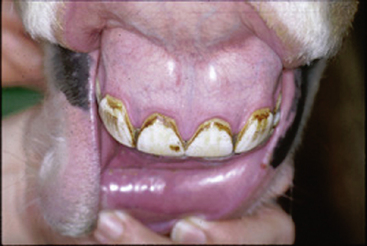
Cyanosis is rare in the horse. Central cyanosis (detectable in mucous membranes) is most commonly due to severe respiratory disease, resulting in hypoxia, or congenital cardiac or circulatory conditions causing right-to-left shunting of blood, thereby bypassing oxygenation in the lungs (Figure 13-2). Central cyanosis is generally not detectable until PaO2 is below 40 mm Hg, below the 60 mm Hg level defined for hypoxia. Evaluation of the response of the PaO2 to nasal insufflation with 100% oxygen, if available, can help indicate whether the cyanosis is due to a right-to-left shunt. A PaO2 of less than 100 mg Hg after nasal insufflation with 100% oxygen is strongly suggestive of a right-to-left shunt.5
The clinical presentation of neonatal foals with a ventricular septal defect (VSD) ranges from normal to debilitated based on the size, location, and number of defects, and the presence of any additional cardiac abnormalities. Small defects may have no gross clinical manifestations, and the defect may go unnoticed until the horse reaches training age or beyond. Typical findings on auscultation of an isolated VSD are a grade 3/6 or higher, harsh, band-shaped pansystolic murmur with the point of maximal intensity over the right third to fourth intercostal space (tricuspid valve region) and a grade lower crescendo-decrescendo ejection-type murmur, with the point of maximal intensity over the pulmonic valve region. The right-sided murmur represents flow through the septal defect, and the left-sided murmur represents the relative pulmonic stenosis that occurs secondary to the right ventricular volume overload created by the defect (i.e., more blood than normal flowing through a normal pulmonic valve). A left-sided murmur that is louder than the right-sided murmur suggests that the septal defect is located in the right ventricular outflow portion of the septum or that other abnormalities exist.6,7 Diastolic murmurs may also exist in conjunction with the typical systolic murmurs of a septal defect and are usually due to prolapse of an aortic leaflet into the defect, and resulting aortic regurgitation.
The clinical presentation of foals with other types of congenital cardiac diseases can also vary widely, ranging from normal to recumbent, hypoxic, or cyanotic depending upon the type and severity of the lesion. Typically, however, foals with complex congenital cardiac disease are severely debilitated.1,4,6 Most foals with congenital cardiac disease demonstrate grade 3/6 or higher holosystolic or pansystolic murmurs. Care should be taken to not equate the loudness or grade of the murmur with the severity of the suspected disease process, as complex congenital abnormalities may have murmurs that are a lower grade than those of a VSD. Likewise, the absence of a detectable murmur should not rule out congenital cardiac disease in cases when it would otherwise be suspected.
PATHOGENESIS
Physiologic flow murmurs are murmurs of left ventricular ejection, due to nonlaminar flow in the great vessels, as in adult horses. Echocardiographic examinations of foals with murmurs that are consistent with normal physiologic flow murmurs have indicated that there is often no other echocardiographically detectable cause for the murmur, such as valvular regurgitations, shunts, or ductal flow.1 Murmurs due to flow in patent remnants of fetal circulation (ductus arteriosus or foramen ovale) can also be normal in the neonate, if not detected beyond the normal time window for closure.
In utero, the fetus receives oxygen via the placental blood flow and the fetal lungs are bypassed, in essence a right-to-left shunt. Fetal circulation bypasses the lungs via the ductus arteriosus, which forms a communication between the pulmonary artery and aorta, and the foramen ovale, which forms a communication between the right and left atria. At birth, decreases in pulmonary resistance and increases in oxygen content associated with inflation of the lung stimulate physiologic closure of the ductus and the foramen ovale. Anatomic closure follows.1,8,9 Physiologic closure of the ductus typically occurs within 24 to 72 hours. Thus, continuous or systolic murmurs within that time frame are considered nonpathologic. Occasionally, systolic flow persists in the ductus for up to several weeks postbirth. This may be a normal variation or secondary to disease processes that result in sustained elevation in right-sided pressures or hypoxia (pneumonia, pulmonary hypertension, dysmaturity/immaturity, maladaptive foal syndrome, or other pulmonary disease). However, systolic murmurs associated with ductal flow should not persist beyond seven weeks of age. Functional closure of the foramen ovale has been suggested to be associated with elevation of left atrial pressures secondary to ductal closure, as well as changes in oxygen tension. Anatomic closure of the foramen ovale occurs during the first few weeks of life.1,9
The true incidence of congenital cardiac abnormalities in the horse is unknown although relatively rare overall.6,10 In an evaluation of approximately 2500 foals, fetuses, and horses presenting for necropsy over four years, Rooney and Franks found four congenital cardiac anomalies.11 Cardiac defects accounted for 3.6% of the abnormalities in a survey of congenitally abnormal fetuses and foals. Marr reports detecting congenital abnormalities in 3.4% of 380 horses presenting for cardiac evaluation.1,12 VSDs are recognized as the most common type of congenital cardiac abnormality in the horse, as in cattle and llamas.1,4,6,23,24 However, a variety of congenital and complex congenital cardiac defects have been described in foals including atrial septal defect, patent ductus arteriosus, VSD with pulmonic stenosis, tetralogy of Fallot, endocardial cushion defect, tricuspid atresia, truncus arteriosus, pseudotruncus arteriosus, transposition of the great vessels, and double outlet right ventricle, among others.1,4,6,10,13–26 Other congenital defects, such as an anomalous origin of the right coronary artery, have been echocardiographically detected in the adult horse and are unlikely to be of clinical significance in the neonate.27
Congenital cardiac abnormalities represent either persistent remnants of fetal circulation after birth (patent ductus arteriosus or patent foramen ovale) or failure of a step or steps during embryologic development of the heart. Membranous VSDs are a result of failure of fusion of the muscular septum with the endocardial cushion during early fetal development, and right ventricular outflow septal defects are a result of abnormal development of the bulbus cordis.4 Defects may also occur in the muscular portion of the septum. VSDs may be present as part of more complex congenital cardiac disease.
The cause of VSD is unknown although evidence suggests it is a heritable defect in cattle and dogs.28 Certain breeds of horse, including Standardbreds, Arabians, American Miniatures, and Welsh Mountain ponies, appear to have an increased incidence of congenital cardiac disease.1,4,29,30 Using horses with VSDs or other congenital cardiac abnormality for breeding purposes is not recommended. Genetic defects, maternal infection, nutrition, and hypoxia have been implicated in small animals and suggested as a cause in large animals.12,19,31,32 Exposure to teratogens in early gestation could potentially play a role in causing abnormal cardiac development in utero, but little evidence exists in this regard.
DIAGNOSTIC TESTS
The diagnostic test of choice for evaluation of a murmur or suspected cardiac disease is an echocardiographic examination using B-mode (two-dimensional real-time) ultrasound, M-mode ultrasound, pulsed and/or continuous-wave Doppler, and color-flow Doppler examination.1,4,6,19,29,31–34 Thoracic radiographs may be useful in suggesting gross cardiac enlargement and vascular changes, but are nonspecific. Likewise, electrocardiographic changes are nonspecific in the equine. Rarely, more invasive techniques such as angiography may be of additional diagnostic benefit. However, they are generally performed when indicated following an initial echocardiographic examination, are limited by the animal’s size (less than 300 pounds), and are generally performed at referral centers.
Stay updated, free articles. Join our Telegram channel

Full access? Get Clinical Tree


