10 Neurologic Dysfunctions
Case 10-1 Foal with Hypoxic Encephalopathy
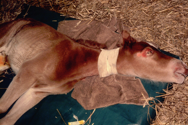
It took the veterinarian approximately one hour to arrive at the farm. During that time, ’05 Carpe Diem became recumbent and was seizing with his head thrown back in opistonus (Figure 10-1). The veterinarian administered 5 mg of diazepam and immediately referred the foal to a neonatal clinic for evaluation. Upon arrival to the clinic, the foal was somnolent and unresponsive. His heart rate was within normal limits, while his temperature was slightly elevated at 102.4° F. Though the foal’s respiratory effort was normal, he appeared to have developed an apneustic (breath-holding) breathing pattern. Mucous membranes were pink with a capillary refill time of <2 seconds. The foal had multiple superficial abrasions around his eyes. A limited neurologic examination was performed because of the foal’s mental status. During the examination, he began to show seizurelike activity manifested by extensor rigidity with opisthotonus and tonic/clonic marching activity.
NEUROLOGIC EXAMINATION IN THE NEONATAL FOAL
The goal of the neurologic examination in the neonatal foal is the same as that of the neurologic examination of the adult horse—lesion localization. Once the lesion is localized, possible etiologies can be explored. There are some unique differences in neurologic response in the neonate and the adult horse. Observation of any deviation from normal behavior patterns or timetable of behavioral events should be noted. As seen in Chapter 1, the newborn foal should assume sternal recumbency soon after birth, stand within two hours of birth and nurse within three hours of birth. The foal should be mentally alert during this time. Foals may exhibit submissive behavior when approached by a human or a horse besides its dam. Submissive behavior in the foal consists of raising its head and exhibiting a chomping motion with its mouth.1 Deviations from a normal behavior pattern can be the result of many different origins, including perinatal asphyxia, intrauterine infection, prematurity, and congenital disease.2,3,4
Upon examination of the cranial nerves of the newborn foal, several differences should be noted. The newborn foal lacks a menace reflex. This is a learned reflex and generally develops over the first two weeks of life. The pupillary light reflex in foals may be affected by their level of excitement perhaps secondary to increased sympathetic tone, with the excited foal having a slightly slower constriction. This may be hard to evaluate in a struggling foal because they will not hold their heads still long enough for the examination. Foals normally have exaggerated head movements in response to external stimuli. The pupils of the foal appear to have a slight ventromedial deviation up until one month of age. Effective nursing generally indicates the normal functioning of cranial nerves 5, 7, 9, 10, 11, and 12.
In evaluating laryngeal function, it has been found that foals do not respond consistently to the “slap test” until about one month of age.3 The test is performed by gently slapping the foal on the withers with a hand while either palpating the larynx or viewing it with an endoscope. A positive test would be the adduction of the arytenoid on the opposite side of the body from the slap. Foals may nicker or whinny right after birth, which is a sign of an alert foal.1 It is not uncommon for foals to protrude their tongues to one side of their mouth or to suck on them. This is particularly seen in foals that have not been allowed to suckle or are orphaned. Tongue tone is normal in these foals and not a result of hypoglossal nerve damage. If the tongue lacks tone, then a neurologic deficit should be suspected in this nerve.
Gait analysis of the foal can be done as the foal moves freely about the stall or paddock. Restraint of the foal may actually get unexpected results. Many foals will collapse in the handler’s arms as restraint is applied.1–3 One’s first reaction is to tighten the restraint and hold the foal up. Though it is counterintuitive, the actual lightening of restraint will cause the foal to bounce up and regain its normal standing posture. Foals will often assume a basewide stance. The newborn foal is obviously clumsy but develops a more coordinated gait within hours of birth. The gait tends to be choppy and dysmetric at first, but as the foal exercises more the gait assumes the more normal gait of the adult.1–3 In foals that have been confined to a stall, the development of the more adult gait is delayed.
Spinal reflexes can be assessed in the recumbent foal. When manipulating the foal’s limbs, one may encounter a strong extensor rigidity that should relax with continued passive motion. Limb reflexes are usually hyperreactive in the foal as compared to the adult. A crossed extensor reflex is commonly seen in the contralateral limb in response to the withdrawal reflex and may be present for the first three weeks of life.1
The suckle reflex is present in all normal foals and is never lost unless a disease process interferes.6 Normal foals can be seen curling their tongue and making suckle movements with in 10 minutes of birth. Suckling is instinctual and though the hypoglossal nerve is responsible for muscular coordination of tongue movement, seeking of the udder and actively suckling is probably under cerebral control as well. Loss of suckle is often the first neurologic sign that an owner may note, and it is usually the last sign to remain as the neurologic syndrome resolves.
Seizures are also a sign of cerebral dysfunction. They are caused by aberrant electrical activity in the brain. It is felt that foals may have a lower seizure threshold because of cortical immaturity.3 Changes in the neuronal membrane, local tissue trauma, or systemic illness can also lower the seizure threshold further.5 Seizures are often described as focal, subtle, or generalized clinical presentations that are dependent on the location and extent of the electrical activity. Subtle or focal signs in the foal may present as oral movements such as random chewing, suckling, or flemen. Rapid eye movements, muscle fasciculations, and apneustic breathing may be present.3 More generalized clinical signs include thrashing, paddling, opisthotonus, extensor rigidity, and hyperthermia. Abrasions to the head and corneal ulcerations are common sequela to an uncontrolled generalized seizure.3
Spinal ataxia can also be seen in the newborn foal but is a less recognized problem than cerebral disease. If present at birth or soon after, then congenital malformations of the spine or spinal cord, vascular accidents during the birth process, and spinal trauma should be considered in the differential diagnosis.2
DIFFERENTIAL DIAGNOSIS FOR SEIZURE IN THE NEONATAL FOAL
Neonatal Encephalopathy
Neonatal encephalopathy (NE), also known as neonatal maladjustment syndrome, hypoxia ischemic encephalopathy, hypoxic encephalopathy, dummy foals, barker foals, and convulsives, is the most common cause of acquired seizures in the foal. In one retrospective study, 18% of foals presented to a neonatal referral center with neonatal maladjustment syndrome.7 Affected foals may present with neurologic dysfunction immediately at the time of birth, or the clinical signs may have a delayed onset but usually within the first 24 hours of life. Hess-Dudan makes the distinction between these two groups of foals based on the onset of clinical signs. Foals that have a delayed onset of clinical signs are usually full-term, have a rapid, seemingly uncomplicated parturition, and have relatively normal postnatal behavior immediately after birth. They may stand and walk and may or may not have an effective suckle. Onset of neurologic signs is usually within six hours. Because the signs are not present immediately at birth, it is felt that the asphyxial trauma to the foal takes place during or immediately after the birth process. The progression of signs is variable, but in general foals lose their suckle reflex, wander away from their dam, become recumbent, and then begin seizing. Not all foals follow this entire process. Signs may only progress to the loss of suckle reflex. This variability in clinical sign progression may be related to the degree of perinatal insult in the foal.8 In human neonatology, the degree of asphyxia (mild, moderate, and severe) is linked to metabolic acidosis with a base deficit >12 mmol/liter.9
Hess-Dudan’s second category of foals with neonatal encephalopathy includes foals that exhibit abnormal behavior immediately after birth. The delivery and/or the placenta may have been abnormal. The neurologic signs may be similar to the first category of foals, but in addition these foals are weak and may show signs of prematurity. It is felt that these foals have experienced a more chronic insult in utero perhaps due to placental insufficiency.8
The etiology of NE is not simple. It is felt that an asphyxial event plays an important part in the process. This event could be secondary to chronic placental insufficiency or acute placental separation. Impaired oxygen delivery results in asphyxia, hypoxia, and sometimes ischemia. In times of low blood oxygen, the fetus tries to maintain blood flow to the brain and heart by redistributing it from the gut, kidneys, liver, and muscle. Brain injury occurs when this mechanism is insufficient. Though not directly studied in foals, studies in neonates of other species (rats, piglets, monkeys, and humans) show that neuronal cell death can be acute from high levels of neurotoxins such as glutamate (produced in response to the asphyxia) and influx of calcium into neuronal cells, or can be delayed as a result of reperfusion injury and release of oxygen radicals.9–14 Reperfusion injury becomes evident hours after the original insult. This may explain why many foals with NE appear normal at birth but begin showing signs of neurologic compromise three to six hours after birth.
The neonate is adapted to handle short nonprogressive periods of hypoxia. Studies looking at a single continuous partial asphyxia in fetal monkeys near term demonstrated brain swelling with neuronal injury.9 For neuronal injury to be sustained, the hypoxic insult must be progressive over a period of time. Injury has been produced by maintaining the hypoxic event for 60 minutes.9 Possible parturition events that might result in progressive hypoxia that would meet this time criteria includes premature separation of the placenta during parturition, umbilical cord compression or torsion, and chest compression in the birth canal for prolonged periods of time.
Postmortem examination of affected foals may or may not be rewarding. Significant vascular accidents (SVA) comprising subarachnoid, parenchymal, and nerve root hemorrhages; cerebral edema; and ischemic necrosis have been described on postmortem examination of some foals that died subsequent to showing signs of neurologic disease.15–18 High vascular pressures that peak at 400 mm Hg during delivery have been hypothesized as a possible cause of the SVA.19
There are many clinical manifestations of neonatal encephalopathy. The respiratory centers are a common target. Abnormal respiratory patterns seen include central tachypnea (midbrain lesion), apneusis (pontine lesion; breath-holding inspiratory pause with no expiratory pause), periodic apnea (midbrain lesion; defined as >20 seconds between breaths), cluster breathing (high medullary lesion; several rapid breaths followed by a respiratory pause or apnea), ataxic breathing (medulla; irregular timing of breaths) and Cheyne-Stokes breathing. These abnormal breathing patterns may or may not be accompanied by central hypercapnia. It is important to detect and treat hypoventilation as it occurs in the early hospital course. Hypercapnia must be detected by blood gas analysis, as often the abnormal respiratory patterns are not accompanied by changes in PaCO2.20 Though the central nervous system signs are the most prominent in cases of perinatal asphyxia, other systems are also affected. In human infants, mild asphyxia may affect systems such the cardiovascular, respiratory, and renal systems mildly or not at all. Moderate to severe asphyxia is more likely to have moderate to severe effects on these systems.8 Renal compromise in the foal with NE may present as decreased urine production. Gastrointestinal effects of perinatal asphyxia may include colic, transient ileus, and necrotizing enterocolitis. Cardiac arrhythmias, edema, and hypotension may result from hypoxia of the myocardium.16
Bacterial Meningitis
Bacterial meningitis is an infrequent manifestation of septicemia in the neonatal foal.21 Koterba found that approximately 10% of foals with confirmed septicemia presented with bacterial meningitis.22 Foals usually have a history of failure of passive transfer and present with the signs of sepsis (see Chapter 5). Neurologic signs of depression, weakness, and anorexia must be distinguished from the generalized septic process in the foal and a more specific localization of meningitis. More specific neurologic signs that may suggest inflammation of the meninges would include cervical pain, proprioceptive deficits, intention tremors, opisthotonus, and coma. It is difficult to distinguish bacterial meningitis from NE because the signs are similar and sepsis often is present in the foal with NE.
The bacteria involved in bacterial meningitis are the same that are involved in sepsis—E. coli, Klebsiella sp., Actinobacillus sp., Salmonella sp., Enterobacter sp., Streptococcus sp., Staphylococcus sp., and Pseudomonas sp. Bacteria gain entry to the meninges by the hematogenous route.2 The central nervous system of the neonate has an incomplete blood-brain barrier. Open tubulocisternal endoplasmic reticulum components of the cerebral endothelial and choroids plexus epithelial cells are thought to be sites of bacterial seeding in the neonate.23 Once bacterial entry has occurred, the cerebral spinal fluid acts as a good media for bacterial growth. Rapid multiplication of the bacteria is favored because of the inadequate humeral and phagocytic defenses of the CNS.23
Inflammation secondary to the activation of the cytokine cascade is a large component of the course of the disease. Brain edema and increased cerebral blood flow initially increase intracranial pressure. As the disease progresses, intracranial pressure increases resulting in a decrease of cerebral blood flow. Ischemia results in irreversible neuronal cell damage.22,23
Trauma
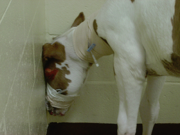
Head and neck trauma in the foal is fairly common in the history of foals with neurologic disease. Generally the history will present a foal that was clinically normal up until the incident of injury. Clinical signs are dependent on the severity and location of the trauma. Cerebral trauma will have signs of blindness, depression, seizure, head pressing and coma (Figure 10-2). Trauma to the brain stem may be accompanied with signs of nystagmus, facial paralysis, strabismus, and gait abnormalities.2,3 Neck trauma results in signs of ataxia or paralysis.25 Fractures of the frontal and parietal bones, the petrous temporal bone, and the basisphenoid bones are common. Intracranial hemorrhage can be parenchymal, subdural, or both. Subdural hematoma formation usually develops from bleeding by a damaged cortical artery or underlying parenchymal injury, or from tearing of a bridging vein form the cortex to a draining venous sinus. Small subdural hematomas often resolve spontaneously, but continued bleeding and expansion of the hematoma leads to increased intracranial pressure and deformation of the brain.26
Congenital Neurologic Problems
Several congenital neurologic problems have been noted in foals, particularly in Arabian foals. Benign pediatric epilepsy has been seen in Arabian and cross Arabian foals particularly from the Egyptian line. Foals are generally normal between seizures. Seizures are unpredictable and may be single or multiple in occurrences. Central blindness for an extended amount of time may be noted as a postictal sign. Foals will often have evidence of trauma around the head. Foals appear to grow out of this problem.3,27
Stay updated, free articles. Join our Telegram channel

Full access? Get Clinical Tree


