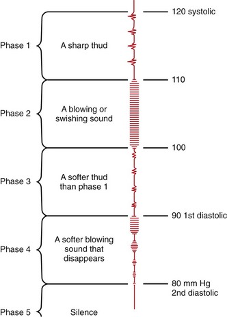
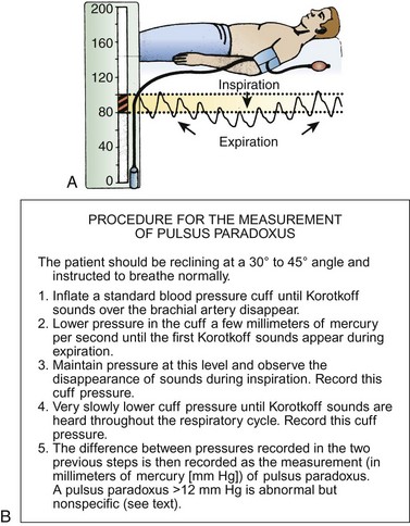
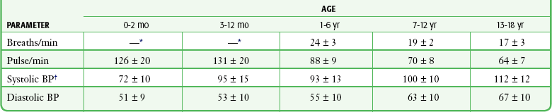
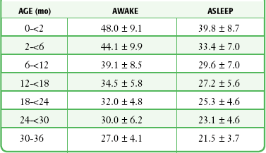
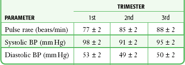
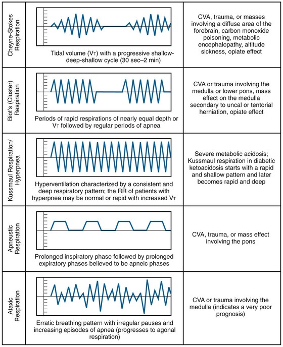
Chapter 1 Measuring the temperature, pulse, respiratory rate (RR), blood pressure, and pulse oximetry is generally recommended for all emergency department (ED) patients, in addition to assessment of pain in the appropriate patient population. For very minor problems or for some fast-track patients (e.g., suture removal), a full set of vital signs may not be required, but this is best decided on a case-by-case basis rather than by strict protocol. Vital signs may not only indicate the severity of illness but also dictate the urgency of intervention. Although a single set of abnormal values suggests pathology, findings on triage or the initial vital signs may be spurious and simply be related to stress, anxiety, pain, or fear. It would be incorrect and not standard of care to attribute initial triage blood pressure, RR, or pulse rate to specific pathology or to retrospectively assume that diagnostic or treatment interventions should have been initiated based solely on these readings. The greatest utility of vital signs, therefore, is their observation and trends over time. Deteriorating vital signs are an important indicator of a compromised physiologic condition, and improving values provide reassurance that the patient is responding to therapy. When a patient undergoes treatment over an extended period, it is essential that the vital signs be repeated as appropriate to the clinical scenario, particularly those that were previously abnormal. In some clinical circumstances it is advisable to monitor the vital signs continuously.1 Vital signs should be measured and recorded at intervals as dictated by clinical judgment or the patient’s clinical state or after any significant change in these parameters. Adhering to protocols or disease categories may not be useful or productive. An abnormal vital sign may constitute the patient’s entire complaint, as in a febrile infant, or it may be the only indication of the potential for serious illness, as in a patient with resting tachycardia.2 Emergency medical service (EMS) personnel begin assessment of the patient’s status and vital signs in the prehospital setting. Surges of epinephrine and norepinephrine commonly occur during transport by the EMS, and these hormones are known to alter vital signs and lead to increases in the heart rate of greater than 10%.3 Vagal influences may also influence EMS-derived vital signs. Prehospital vital signs should always be interpreted with the entire clinical scenario in perspective. Early recognition of vital signs dates back to the fourth century bc, when Herophilus first described sphygmology, or palpation of the pulse in terms of size, frequency, force, and rhythm. Chinese clinicians (second century bc) timed the pulse by the RR of the examiner in the belief that four pulsations per respiration was normal for adults. The study of pulses was greatly influenced by Galen, who expanded the subject into a rather complex and obscure art form and wrote 18 books on the subject.4 Blood pressure was first measured directly in 1733 by Hales, who recorded arterial pressure in a mare by cannulation with a brass pipe and a blood-filled glass column.5 Frank used large-bore catheters connected to a rubber membrane in a 1903 manometer.6 Invention of inflatable cuff manometers (Riva-Rocci, 1896) and discovery of the arterial phase sounds (Korotkoff, 1905) allowed the development of indirect measurement of blood pressure.5,6 Clinical thermometry was introduced by Sanctorius in 1625, with early thermometers being filled with alcohol. Mercury column thermometers were introduced by Fahrenheit in 1714. Although their routine use was supported by Boerhaave, thermometry was not established as routine clinical practice until the 1870s.7 Published vital sign norms for children are not as well accepted as those for adult patients. Table 1-1 and Table 1-2 report normal vital signs for children by age group as means and standard deviations. In Table 1-1, the values for pulse and blood pressure for 0- to 2-month-olds are adapted from studies of newborn populations (i.e., <7 days).8–10 During the newborn period, normal arterial blood pressure rises rapidly. Values for pulse and respiration in children older than 3 years reflect an average of male and female values for 0- to 1-, 3-, 9-, and 16-year-old populations. The values for blood pressure reflect an average of male and female values for the 1- to 6-month-old and the 3-, 9-, and 16-year-old populations.11 Newer studies have reassessed the reference values for RR in children.12–14 Table 1-2 reflects age-related changes and the effect of the state of wakefulness on RR in children up to 3 years of age.14 A study evaluating resting RRs in pediatric ED patients up to the age of 18, however, indicated considerable patient variability and somewhat higher RRs than those shown in Table 1-2.12 TABLE 1-1 Normal Values for Vital Signs in Infants and Children (Means ± SD) BP, blood pressure; SD, standard deviation. *For data on children 0 to 36 moths, see Table 1-2. †As an estimate, for children 1 to 10 years of age, (2 × age [yr]) + 90 mm Hg = 50th percentile for systolic BP. TABLE 1-2 Normal Respiratory Rates (Breaths/min) in Children Up to 3 Years of Age (Means ± SD) Adapted from Rusconi F, Castagneto M, Gagliardi L, et al. Reference values for respiratory rate in the first 3 years of life. Pediatrics. 1994;94:351. For the adult population, normal blood pressure values have been established. Although systolic blood pressure increases with age, normotensive or normal systolic blood pressure is defined as 90 to 140 mm Hg, and normotensive or normal diastolic blood pressure is defined as 60 to 90 mm Hg. The recent literature suggests defining an “optimal” blood pressure as 115/75 because values at or below this level have been associated with minimal vascular mortality.15 It has been suggested that the definition of hypertension be further expanded to integrate a global cardiovascular risk assessment.16,17 Although most patients have similar blood pressure in both arms, Pesola and coworkers found that 18% of their hypertensive population18 and 15% of their normotensive population had a difference of greater than 10 mm Hg in systolic blood pressure between arms.19 Within the adult population, optimal definitions for normal systolic blood pressure probably vary by age, and particular differentiation should be made in regard to geriatric patients in the emergency setting. The recent literature suggests redefining values representative of hypotension in the elderly, especially in the setting of trauma. Systolic blood pressure readings ranging from approximately 90 to 120 mm Hg have been associated with occult hypoperfusion and increased mortality in geriatric trauma patients.20–22 In 1928 the New York Heart Association, by consensus, established the normal limits for the resting heart rate as 60 beats/min and 100 beats/min. More recent data indicate that 45 beats/min and 95 beats/min may better define the heart rate limits of normal sinus rhythm in adults of all ages. Spodick recommended that the operational definition for the limits of the resting heart rate in adults be 50 beats/min and 90 beats/min.23,24 This view is widely supported among cardiologists,25,26 but these ranges have not been validated in the ED setting. There is currently no consensus on what constitutes a normal adult RR; however, an RR range of 12 to 24 breaths/min is generally accepted in the existing literature as the norm for adults.27,28 Pregnancy results in alterations in the normal adult values for pulse and blood pressure. The RR is unchanged, although the physiologic hyperventilation of pregnancy is well recognized and results from increased tidal volume and decreased residual and expiratory reserve volume.29 The resting pulse rate increases throughout pregnancy to 10% to 15% over baseline values. The norms for systolic and diastolic blood pressure are dependent on patient positioning. When a pregnant patient is sitting or standing, systolic pressure is essentially unchanged. Diastolic pressure declines until approximately 28 weeks’ gestation, at which time it begins to rise to nonpregnant levels. When a pregnant patient is in the lateral decubitus position, both systolic and diastolic pressure declines until the 28th week and then begins to rise to nonpregnant levels (Table 1-3).30 TABLE 1-3 Vital Signs during Pregnancy in the Lateral Decubitus Position (Means ± SD) BP, blood pressure; SD, standard deviation. Adapted from Katz R, Karliner JS, Resnik R. Effects of a natural volume overload state (pregnancy) on left ventricular performance in normal human subjects. Circulation. 1978;58:434. By permission of the American Heart Association. Breathing is initiated and primarily controlled in the medullary respiratory center of the brainstem. The respiratory center is modulated by the pneumotaxic center, which limits the length of the inspiratory signal and greatly influences the RR and apneustic center in the pons.31 Respiratory frequency reveals only a glimpse of the entire clinical picture. The pattern, effort, and volume of respiration may be more indicative of altered respiratory physiology. An abnormality in respiration may be a primary complaint or a manifestation of other systemic diseases. Increased RRs may be seen in patients with a variety of pulmonary or cardiac diseases, but acidosis, anemia, temperature, stress, and drugs (such as stimulants and salicylates) can significantly alter the RR in the absence of cardiopulmonary dysfunction. The respiratory status of both adults and children plays a crucial role in determining the overall assessment of illness. Although it is a sensitive yet nonspecific indicator of respiratory dysfunction, the RR can also predict nonpulmonary morbidity. Several prehospital and hospital-based illness or injury severity scores feature the RR as a cardinal value. A prehospital RR of less than 10 or greater than 29 breaths/min is associated with major injury in 73% of children.32 Using tachypnea alone as a predictor of pulmonary pathology, infants with an RR higher than 60 are found to be hypoxic 80% of the time.33 Pediatric studies have linked abnormal RRs to in-hospital mortality and the level of care required in the ED.34,35 In a retrospective study exploring predictors of critical care admission for adult ED patients who were initially triaged as having low to moderate acuity, an abnormal RR at the first nursing assessment increased the odds of critical care admission by a factor of 1.66.36 An RR higher than 25 breaths/min in prehospital trauma patients was associated with increased mortality.37 Pre-arrest respiratory insufficiency (RR >36 breaths/min or pulse oximetry <90%) was an independent predictor of mortality (odds ratio [OR], 4.2) in patients with EMS-witnessed cardiac arrest.38 Although some studies have associated abnormal RRs in adult ED patients with increased mortality,39,40 a recent large prospective cohort study of adult patients found that an initial abnormal RR on triage in the ED was not an independent predictor of hospital mortality.41 To measure RR (inspirations per minute), count the respirations when the patient is unaware that his or her breathing is being observed. Count for a full minute to most accurately determine the RR. The frequency of breathing is less regular than the pulse, and inaccurate measurement is more likely to occur if the count is taken for a shorter interval. An infant’s RR can easily be determined by observing or palpating the excursion of the chest or the abdominal wall.42 Infants should be observed for grunting respirations, which are produced by expiration against a partly closed glottis (an attempt to maintain positive airway pressure). The reproducibility of RR measurements may be limited by significant interobserver variability.43,44 Clinicians should recognize this inherent variability and interpret the RR with caution. Rates obtained by nurses versus medical students varied significantly, as did those obtained by medical students versus residents and attending clinicians.45 Interobserver variability may account for a difference of up to 6 breaths/min, and variability in the same observer may account for up to 5 breaths/min.45 A study comparing RRs obtained by triage nurses with an electronic monitor found that neither provided an accurate measurement of the RR in the ED, thus suggesting that new clinical strategies for obtaining this vital sign may be necessary.46 Current texts vary considerably in their definition of a normal RR and cite published values that range from 8 to 24 breaths/min. In a study that specifically investigated normal RRs in an ED (afebrile ambulatory patients without respiratory complaints), females had a mean RR of 20.9 breaths/min and males had a mean RR of 19.4 breaths/min. The researchers concluded that a normal RR in the adult patient population was 16 to 24 breaths/min.45 Other studies have provided additional information on normal resting and sleep-state RRs in children younger than 7 years.9–14 RRs obtained with a stethoscope were higher than those obtained by observation (mean difference, 2.6 breaths/min in awake and 1.8 breaths/min in asleep children). Smoothed percentile curves demonstrated a larger dispersion at birth (5th percentile, 34 breaths/min; 95th percentile, 68 breaths/min), whereas dispersion was less at 36 months of age (5th percentile, 18 breaths/min; 95th percentile, 30 breaths/min). The RR will generally increase in the presence of fever. It is often difficult to determine whether tachypnea is a primary finding or is simply associated with hyperpyrexia. A study of children younger than 2 years in whom pneumonia was subsequently diagnosed found that age-appropriate limits for resting tachypnea in the presence of fever could be defined. A sensitivity of 74% and specificity of 77% for pneumonia were found when children 6 months of age had an RR higher than 59 breaths/min, when those aged 6 to 11 months had an RR higher than 52 breaths/min, and when those 1 to 2 years old had an RR higher than 42 breaths/min.47 Even in the face of physiologic compensation for fever, interpretation of the RR alone can help predict the presence of pulmonary disease. Hyperventilation and hypoventilation can result from an extensive variety of disorders and may be related to pulmonary or extrapulmonary pathology. Abnormal respiratory patterns can be characteristic of metabolic or central nervous system pathologic conditions (Fig. 1-1) and may aid in the differential diagnosis. Kussmaul respirations describe the hyperventilation pattern seen in diabetics with ketoacidosis. Hyperpnea, or a normal RR but clinically significant hyperventilation secondary to increased tidal volume, may be seen with salicylate poisoning.48 Figure 1-1 Abnormal respiratory patterns. CVA, cerebrovascular accident; RR, respiratory rate. (Adapted from Breathing Patterns. © 2011—D’Urbano J—Breath Sounds. Available at http://www.BreathSounds.org.) Observe the respiratory patterns carefully in children. In infants it is essential to distinguish normal periodic breathing from apnea. By definition, periodic breathing consists of three or more respiratory pauses longer than 3 seconds in duration with less than 20 seconds between pauses. There is no associated bradycardia or cyanosis. This contrasts with apnea, which is a particular problem in preterm infants. Apnea is defined as a respiratory pause longer than 20 seconds. It may be associated with bradycardia and hypoxia.42 Periodic breathing and apnea are believed to be disorders on a continuum, both stemming from abnormal physiologic control of respiration. Periodic breathing is considered a benign disorder, but infants with symptomatic apneic episodes that result in apparent life-threatening events are thought to be at increased risk for sudden infant death syndrome.49 Assessment of blood flow by palpation of the pulse can be used to gauge the presence of cardiac contractility and not just the electrical rhythm. Caution should be taken to not over-generalize the presence or strength of a pulse in predicting blood pressure. The necessity for repeated pulse evaluations is dictated by the clinical complaint and the status of the patient. Continuous monitoring is not routine but may be helpful when the clinical situation may predict significant variability in heart rate, as in the setting of sepsis.50 An association between absence of a radial pulse or absence of both radial and femoral pulses and hypotension has been demonstrated in hypovolemic trauma patients. The variability in individual response prohibits the use of this parameter as an absolute gauge of blood pressure.51 Depending on the clinical scenario, pulses are palpable at numerous sites, although for convenience the radial pulse at the wrist is routinely used. Use the tips of the first and second fingers to palpate the pulse. The two advantages of this technique are that (1) the fingertips are quite sensitive, thereby enabling the pulse to be located easily and counted, and (2) the examiner’s own pulse may be erroneously counted if the thumb is used instead of the first and second fingers. Pulses are also easily palpated at the carotid, brachial, femoral, posterior tibial, and dorsalis pedis arteries. Palpate the pulse at the brachial artery to appreciate its contour and amplitude. Locate the pulse at the medial aspect of the elbow and note that it is more easily palpated when the elbow is held slightly flexed.52 Determine the pulse rate by counting for 1 minute, particularly if any abnormality is present. Common convention in the acute care setting is to count a regular pulse for 15 seconds and multiply the resulting number by 4 to determine the beats per minute. In newborns, use direct heart auscultation and umbilical palpation as the methods of choice to determine the heart rate. Instantaneous changes in newborn heart rates are best indicated to the resuscitation team by the clinician tapping out each heartbeat.53 In unstable children, palpate the central arteries, particularly the femoral and brachial pulses, instead of the more peripheral arteries. In a comparison of four methods of determining the heart rate in infants, listening at the apex of the heart was found to be more accurate than palpation of the brachial, carotid, or femoral pulses.54 Of the sites for palpation of the heart rate, the femoral artery has proved most valuable, especially in hypotensive infants.55 Consider the individual’s physiology when interpreting the pulse. In infants and children, interpret the pulse rate with reference to age (See Table 1-1). Pulse varies with respiration: it increases with inspiration and slows with expiration. This is known as sinus dysrhythmia and is physiologic. Although bradycardia is defined as a heart rate lower than 60 beats/min in adults, a well-conditioned athlete may have a normal resting heart rate of 30 to 40 beats/min.56,57 As discussed earlier, a redefinition of bradycardia to less than 45 beats/min and tachycardia to greater than 95 beats/min has been proposed based on a normal healthy population.24,58 Such definitions include 95% of the population and do not address any given individual’s normal baseline rate. In addition to determining the pulse rate, obtain information about the regularity of the pulse by palpation. An irregular pulse suggests atrial fibrillation or flutter with variable block, and accurate assessment of the pulse should be done by auscultation of the apical cardiac sounds. The apical pulse is frequently greater than the peripheral pulse because of inadequate filling time and stroke volume, with resultant nontransmitted beats. A greater pulse deficit generally reflects more severe disease.59 Accurate examination and description of pulse amplitude and contour can provide additional clinical information and aid in decision making. Superimposition of one pathophysiologic state on another may modify the pulse. For example, sepsis may result in variable pulse amplitudes, depending on the stage in the development of the disease at initial evaluation of the patient. Early in sepsis, cardiac output increases and vascular resistance decreases, which causes bounding pulses. In advanced sepsis or septic shock, falling cardiac output and increased vascular resistance are seen, and pulses are diminished.60 Definable age-related changes in pulse amplitude and contour can be identified. Such changes are due to an increase in arterial stiffness, which results in increased pulse wave velocity and progressively earlier wave reflection. This leads to increased pulse amplitude in the elderly at all commonly measured sites (carotid, femoral, and radial).60 In addition to these age-related changes, pulse wave analysis may be useful in determining arterial stiffness and the likelihood of atherosclerotic disease in a vascular laboratory setting.61 Weak pulses can be a significant finding in patients with hypotension if present globally or an indication of limb ischemia if isolated to one extremity. Bounding pulses can be seen with a widened pulse pressure, which is discussed later in the section on blood pressure. Routine measurement of pulse amplitude is not reproducible by simple palpation and requires instrumentation not available in EDs. Palpated “femoral pulses” during chest compression may represent either forward arterial blood flow or “to-and-fro” movement of blood from the right side of the heart to the venous system. A carotid pulse is preferred when assessing the adequacy of chest compressions during cardiopulmonary resuscitation (see Chapter 17). Changes in arterial blood pressure over time may indicate success of treatment or worsening of the patient’s overall condition. An abrupt reduction in a patient’s arterial blood pressure usually indicates the need for immediate intervention or reconsideration of therapy. The current section discusses indirect blood pressure monitoring, and intraarterial techniques are considered elsewhere. Discussion of the specific use of the Doppler device for measurement of pulse and blood pressure and for measurement of orthostatic blood pressure and changes in pulse follow this section. Despite an association between the absence of hypotension and a radial pulse or between hypotension and the absence of both radial and femoral pulses, in the setting of trauma, the variability in individual response prohibits the use of this parameter as an absolute gauge of blood pressure.52 Arterial blood pressure indicates the overall state of hemodynamic interaction between cardiac output and peripheral vascular resistance. Arterial blood pressure is the lateral pressure or force exerted by blood on the vessel wall. It indirectly measures perfusion, and blood flow equals the change in pressure divided by resistance. Because peripheral vascular resistance varies, a normal blood pressure does not confirm adequate perfusion.62 Mean arterial blood pressure (MAP) can be estimated by adding one third of the pulse pressure (i.e., the difference between systolic and diastolic blood pressure) to diastolic pressure or by using the following measure.63 Patients with minor ambulatory complaints unrelated to the cardiovascular system may not necessarily need their blood pressure measured in the ED, but those with hemodynamic instability need frequent monitoring of blood pressure. In children there is a significant amount of variability regarding standard situations that require measurement of blood pressure. In general, the younger the patient, the less likely blood pressure will be measured.64,65 In newborns, infants, and even toddlers, capillary refill is sometimes substituted for standard blood pressure measurement, although viewing these tests as equivalent can lead to significant errors. In low-flow states, Doppler measurement of blood pressure may be obtained rapidly. Repeated measurements will provide an evaluation of the adequacy of resuscitation in patients whose blood pressure cannot be auscultated by standard techniques and in those in whom intraarterial blood pressure measurements are either contraindicated or technically unobtainable.66,67 Placing a catheter for direct intraarterial measurement of blood pressure has a higher risk for complications, but it may be performed safely in the ED. In particular, direct measurement of arterial pressure during pulseless electrical rhythms may help discriminate between severe shock and otherwise nonresuscitatable status.68 Alternative noninvasive devices for continuous blood pressure measurement (CBPM) have been introduced clinically, with varying success. One common method of CBPM uses finger cuffs equipped with infrared (IR) photoplethysmography and sophisticated technology for quantification of finger blood pressure levels. Finapres (Ohmeda, Madison, WI) was the first commercial product using this technique, and several newer products are on the market today. A number of commercial systems use an alternative method of arterial applanation tonometry to measure CBPM. Further study is needed for solid validation of devices using these techniques.69,70 The equipment required for indirect blood pressure measurement includes a sphygmomanometer (cuff with an inflatable bladder, inflating bulb, controlled exhaust for deflation, and manometer) and a stethoscope, Doppler device (for auscultation), or oscillometric device.71–74 A common practice in the prehospital and interhospital transport setting is to forego auscultatory blood pressure measurements with a stethoscope and instead obtain systolic values only by palpation of the first Korotkoff sound. This practice, though sometimes the only feasible method of obtaining any value in a noisy environment, poses a significant potential for error. In a study of critically ill patients transferred between hospitals, palpated systolic blood pressure values underestimated manometric values by nearly 30%. According to the American Heart Association guidelines, the sphygmomanometer cuff should be an appropriate size for the patient to ensure an accurate reading. The width of the bladder should be at least 40% of the distance of the limb’s midpoint (i.e., from the acromion process to the lateral epicondyle). This published figure of the ideal width, when studied in a validation review, may be higher, up to approximately 50%.75 The length of the bladder should be 80% of the midarm circumference or twice the recommended width.66 Discrepancies in matching upper arm size with cuff size have been demonstrated to produce significant errors in critically ill populations when compared with invasive intraarterial blood pressure measurements.76 The availability of appropriately sized cuffs appears to be a pervasive problem, especially since about 80% of patients do not fit the standard 12-cm large cuffs.77 In one study, 90% of aneroid devices had only one size of cuff available.78 A second study phase from this group showed no marked improvement in agreement of oscillatory and invasive measurements despite correct cuff size.79 Manometers require annual servicing. Mercury columns may require the addition of mercury to bring the edge of the meniscus to the zero mark. The air vent or filter at the top of the mercury column should also be checked for clogging. An aneroid manometer should be calibrated against a mercury column at least yearly. If the aneroid indicator is not at zero at rest, the device should not be used.80 Digital manometers may not be validated for all patient groups and could give inaccurate readings. Automatic sphygmomanometers may improve physiologic monitoring with their alarm and self-cycling capabilities. They offer indirect arterial blood pressure measurement with little pain and without the risks associated with invasive arterial lines.81 Accuracy of measurements does not suffer during rapid cycling, but the potential for vascular injury from nearly continuous arterial compression dictates that most automated blood pressure units will revert back to less frequent (i.e., every 15 to 20 minute) cycling as a safety precaution. Oscillometric blood pressure monitors detect motion of the blood pressure cuff transmitted from the underlying artery. A sudden increase in the amplitude of arterial oscillations occurs with systolic pressure and MAP, and an abrupt decrease occurs with diastolic pressure.82 In adult patients, numerous studies have focused on the reliability of auscultatory versus automated blood pressure measurements. Mercury column versus Dinamap readings showed increased disparity with systolic blood pressure greater than 140 mm Hg, the range at which accuracy should be most rigorously sought to correctly identify hypertension. In general, automated blood pressure devices yield higher systolic and lower diastolic blood pressure.83 The range of error in automated devices was, on average, 4.0 to 8.6 mm Hg.84,85 Unfortunately, these studies represent populations without critical illness and do not reflect the accuracy of readings at the extremes of hypertension and hypotension, thus making generalization to an ED population difficult.85,86 Obtain indirect blood pressure measurements at the patient’s bedside by palpation, auscultation, Doppler, or oscillometric methods. The technique is straightforward and accurate when the equipment is well maintained, calibrated, and used by clinicians who follow accepted standards. The patient may be lying or sitting, as long as the site of measurement is at the level of the right atrium and the arm is supported.66,74 Unless the arm is kept perpendicular to the body with the elbow resting on a desk, measurements will be 9 to 14 mm Hg higher, regardless of body position.75,87 Allowing the arm to be parallel to the body when supine but supporting the arm perpendicular to the body when measuring blood pressure may create a pseudo-drop in blood pressure. These changes are thought to be dependent on the mechanical properties of the arteries themselves and not associated with hydrostatic pressure alone.88 To palpate arterial blood pressure, inflate the cuff to 30 mm Hg above the level at which the palpable pulse disappears. Once properly inflated, palpate directly over the artery and deflate the cuff at a rate of 2 to 3 mm Hg/sec. Report the initial appearance of arterial pulsations as the palpable blood pressure. This practice, known as the Riva-Rocci palpatory technique, has shown mixed results in yielding accurate estimations of blood pressure. One study determined that the average underestimation of systolic blood pressure was 6 mm Hg.89 Another operative study looking at the combination of palpated systolic blood pressure and observed visual return of continuous pulse oximetry reported an underestimation of 10 to 20 mm Hg.85 The same technique is used with the Doppler device, but the palpated pulse is replaced with the Doppler auditory signal. Measurement of arterial pressure by palpation and Doppler yields only estimates of systolic blood pressure. The Doppler method is preferred when determining blood pressure in infants.86 When auscultating blood pressure at the brachial artery, apply the blood pressure cuff about 2.5 cm above the antecubital fossa with the center of the bladder over the artery.74 Apply the bell of the stethoscope directly over the brachial artery with as little pressure as possible.90 Systolic arterial blood pressure is defined as the first appearance of faint, clear, tapping sounds that gradually increase in intensity (Korotkoff phase I). Diastolic blood pressure is defined as the point at which the sounds disappear (Korotkoff phase V).74–91 In children, phase IV defines diastolic blood pressure (Fig. 1-2).7 Phase IV is marked by a distinct, abrupt muffling of sound when a soft, blowing quality is heard. It is best to measure by auscultation over the brachial artery because of accepted standardization of the measured values. Alternative sites include the radial, popliteal, posterior tibial, or dorsalis pedis arteries, although any fully compressible extremity artery may be used. Studies evaluating direct and indirect blood pressure measurements have demonstrated good correlation between these methods.92,93 It may not be feasible to obtain upper extremity blood pressure measurements because of patient access issues, particularly those encountered in the prehospital setting. Forearm measurements may be obtained more easily and show fair correlation to standard upper extremity values (within 20 mm Hg in 86% of systolic measurements and 94% of diastolic measurements).94 Alternatively, noninvasive finger blood pressure measurements have shown promise when compared with standard upper extremity readings. The overall discrepancy in an ED study was 0.1 mm Hg with a standard deviation of ±5.02 mm Hg when comparing finger blood pressure and invasive MAP via radial artery cannulation.95 Novel noninvasive continuous finger cuff technology offers the benefit of uninterrupted monitoring and has the advantage over invasive techniques of being safer and immediately available. Noninvasive finger cuff measurements have shown reasonable correlation, even in critically ill populations in the ED.70 Wrist blood pressure has shown to have good average accuracy in the surgical environment when compared with oscillometric devices. Patient comfort is reported to be greater with these devices. The typically stated contraindications to the acquisition of upper arm blood pressure (limitation after mastectomy, etc.) may not apply.96 The accuracy of the palpatory, Doppler, and oscillometric methods has also been investigated.97–99 When phase I and V Korotkoff sounds are used, indirect methods typically underestimate systolic and diastolic pressure by several millimeters of mercury.97–100 During shock, the palpatory and auscultatory methods underestimate simultaneous direct arterial pressure measurements.101 The flush method, in which return of color after deflation of the cuff is used to estimate blood pressure in infants, may underestimate systolic blood pressure by up to 40 mm Hg.102 This method is unreliable and not recommended. Complications of indirect blood pressure measurements are minimal when the proper procedure is followed. Inadvertent prolonged application of an inflated blood pressure cuff may result in falsely elevated diastolic pressure and ischemia distal to the site of application.76 Invasive blood pressure monitoring is associated with a number of potential problems (see Chapter 20). Normal blood pressure increases with decreasing distance from the heart and aorta. Blood pressure tends to increase with age and is generally higher in males. Individual factors that influence blood pressure include body posture, emotional or painful stimuli, environmental influences, vasoactive foods or medications, and the state of muscular and cerebral activity. Exercise and sustained isometric muscular contraction increase blood pressure in proportion to the strength of the contraction. A normal diurnal pattern of blood pressure consists of an increase throughout the day with a significant, rapid decline during early, deep sleep.103 Normal lower limits of systolic blood pressure in infants and children can be estimated by adding 2 times the age (in years) to 70 mm Hg. The 50th percentile for a child’s systolic arterial blood pressure from 1 to 10 years of age can be estimated by adding 2 times the age (in years) to 90 mm Hg. Children older than 2 years are considered hypotensive when systolic blood pressure is less than 80 mm Hg.104 Children are able to maintain MAP until very late during shock.105 The finding of a normal blood pressure in a child with signs of poor perfusion should not dissuade the clinician from appropriate treatment. Most adults are considered hypotensive if systolic blood pressure is lower than 90 mm Hg, but some individuals normally exhibit a systolic pressure in that range. In the elderly, the presence of normotension within defined or published limits may not be reassuring. When considering systolic blood pressure cutoffs for trauma patients, 85 mm Hg for patients aged 18 to 35, 96 mm Hg for patients aged 36 to 64, and 117 mm Hg for those older than 65 have been proposed as new standards for hypotension.20 When accompanied by signs of shock, immediate treatment is indicated. In patients with shock, blood flow cannot be reliably inferred from heart rate and blood pressure values.106,107 Adults are hypertensive if either systolic or diastolic pressure consistently exceeds 140 or 90 mm Hg, respectively.108,109 A meta-analysis showed strong correlation of blood pressure to vascular and overall mortality down to at least 115/75.110 Some authors have suggested altering the blood pressure definitions to include an “optimal” blood pressure of 115/75.15 Other authors have suggested incorporating blood pressure into a global cardiovascular risk assessment that includes other associated risk factors.16,17 The applicability of population norms for hypertension in a stressful emergency situation is controversial. One should not make diagnostic or therapeutic decisions based solely on an abnormal initial measurement. Patients with hypertension require repeated measurements to assess whether therapy is required in the ED. Because sustained hypertension may be seen in more than a third of initially hypertensive ED patients, careful evaluation and follow-up are required.111 The phenomenon of white coat hypertension (WCH) is defined as a persistent elevation in blood pressure in the clinical setting only. The prevalence of WCH is between 20% and 94%, depending on the frequency of reassessment of the clinical setting.112 It is unclear whether patients who have isolated hypertension in the clinical setting (WCH) are at increased risk for the development of hypertension and subsequent end-organ damage.113 Erroneous blood pressure measurements may result from several factors.114 Falsely low blood pressure may be caused by using an overly wide cuff, by placing excessive pressure on the head of the stethoscope, or by rapid cuff deflation.115,116 Falsely high blood pressure may be caused by the use of an overly narrow cuff, anxiety, pain, tobacco use, exertion, an unsupported arm, or slow inflation of the cuff.117 There appears to be a statistically significant difference in the error rate associated with patients weighing more than 95 kg, whether from obesity or as a result of muscular upper arms from body building.118,119 Of note, 41% of adults observed at the University of Pittsburgh required non–standard-sized cuffs, and the use of small cuffs was associated with a mean error of 8.5 and 4.6 mm Hg in systolic and diastolic pressure, respectively.116 Other studies have confirmed relatively high rates of inappropriately diagnosed hypertension in obese patients based on erroneous cuff size.118 Other specific study populations in this area have been critically ill patients, in whom disparate cuff size can lead to significant inaccuracies based on arm circumference.82 Hypotensive patients have unreliable Korotkoff sounds, but Doppler measurements are well correlated with direct arterial systolic pressure measurements in hypotensive patients.120 An auscultatory gap can be appreciated in hypertensive patients and may mislead the clinician. It is heard during the latter part of phase I and should not be confused with diastolic readings. Auscultation until the manometer reading approaches zero should prevent misinterpretation. In patients with aortic insufficiency or hyperthyroidism, in those who have just finished exercising, and in children younger than 5 years, measurement of diastolic blood pressure should occur at Korotkoff phase IV. Extremes of blood pressure, both hypotension and hypertension, have been found to be factors contributing to measurement errors in critically ill pediatric patients. Predictably falsely high readings for noninvasive versus invasive measurements have been obtained in hypotensive patients and falsely low values in hypertensive states.121 Hemiplegic patients may exhibit different blood pressure in the affected and unaffected arms.122 A flaccid extremity tends to yield lower systolic and diastolic pressure, whereas a spastic extremity tends to yield higher values than the extremity with normal motor tone. Although these differences are generally small, it is preferable to monitor blood pressure in the unaffected limb. Numerous errors may occur in the accurate measurement of blood pressure. The only way to combat them is to first be cognizant of practices contributing to them. Unfortunately, few nurses can identify causes of potentially erroneous readings. In a study examining nurses’ ability to obtain accurate readings, proper technique in determining systolic blood pressure could be identified 61% of the time; diastolic blood pressure, 71% of the time; and an auscultatory gap, 54% of the time. Nurses were able to correctly identify faulty equipment 58% of the time, assess cuff size 57% of the time, determine appropriate inflation pressure 29% of the time, note the appropriate deflation rate 62% of the time, and determine correct arm positioning 14% of the time.123 With the increasing number of patients with heart failure, those receiving bridging measures to transplantation, or those treated with long-term circulatory augmentation devices in the form of left ventricular assist devices (LVADs), it useful to understand the difficulty in interpreting blood pressure measurements in these patients. All types of VADs fit into two categories: (1) pulsatile and (2) nonpulsatile. Pulse and blood pressure readings in patients with pulsatile VADs (Thoratec, HeartMate IP and VE, Novacor) are comparable to values in the general non-VAD population. Nonpulsatile VADs (Levitronix Centri Mag, Tandem Heart, Impella, DeBakey LVAD, HeartMateII) function by either centrifugal or axial blood flow, and this has a significant impact on the ability to detect pulses.124 Typically, these patients appear to be perfusing well with adequate skin warmth and capillary refill even though pulses may be absent. Blood pressure readings can be obtained with these nonpulsatile flow devices, but diastolic blood pressure, pulse pressure values, and MAP vary significantly depending on the speed of the pump.125 The presence of a systolic blood pressure differences of 10 to 20 mm Hg between the arms suggests a normal condition. If greater, it may indicate advanced focal atherosclerosis, coarctation of the aorta proximal to the left subclavian artery, type A aortic dissection, aortic arch syndromes, or other vascular processes preferentially affecting one extremity. The utility of upper extremity bilateral blood pressure measurements has recently come into question. One study found a 10-mm Hg systolic or diastolic difference in 53% of patients in the emergency setting and a 20-mm Hg or higher difference in 19% of patients.126 Although these numbers have not generally been found to be of this magnitude in metaanalysis,127 the unique setting of the study in the ED makes correlation particularly salient for the emergency physician. The reliability of peripheral pulse deficits in diagnosing or excluding type A aortic dissection is a frequently cited reason for evaluating blood pressure in both arms. In a metaanalysis, Teece and Hogg noted that the absence of a clinical pulse deficit to exclude thoracic dissection in patients with chest pain was just 31%, and the authors concluded that peripheral pulse deficits are far too insensitive to warrant their use as a means of excluding thoracic aortic dissection in patients with chest pain.128 Given that many in the general population have significant differences in blood pressure in each arm, the diagnostic value of this frequently cited indication for obtaining bilateral brachial blood pressure is unproven. Essentially, most patients with a type A aortic dissection will not have a measurable blood pressure discrepancy between the arms, and most of those who do have such a finding will not have dissection. Brachial pressure differences did not appear to be linked to age, gender, race, MAP, cardiovascular risk, or final discharge diagnosis. Smaller interarm differences have been reported in the ED setting (18% in hypertensive patients and 15% in normotensive patients when a cutoff greater than 10 mm Hg was used).18,19 Though not tested, a method proposed to minimize these differences in the ED is to take simultaneous blood pressure readings from both the left and right extremities with two calibrated automated blood pressure units.129 Normal respiration briefly decreases systolic blood pressure by approximately 10 mm Hg during inspiration. Pulsus paradoxus occurs when there is greater a than 12–mm Hg decrease in systolic blood pressure during inspiration. Pulsus paradoxus may occur in patients with chronic obstructive pulmonary disease, pneumothorax, severe asthma, or pericardial tamponade.101 To measure a paradoxical pulse, have the patient lie comfortably in the supine position at a 30- to 45-degree angle and breathing normally in an unlabored fashion (which are unusual conditions in a patient suspected of having cardiac tamponade, severe asthma, chronic obstructive pulmonary disease, or pneumothorax).130 Inflate the blood pressure cuff well above systolic pressure and slowly deflate it until the systolic sounds that are synchronous with expiration are first heard (Fig. 1-3). Initially, the arterial pulse will be heard only during expiration and will disappear during inspiration. Deflate the cuff further until arterial sounds are heard throughout the respiratory cycle. Palpation at the radial or femoral arteries may yield complete disappearance during inspiration. When present, this technique is a quick bedside confirmation of the possibility of severe tamponade. An alternative method for determination of pulsus paradoxus is by visually observing loss of the pulse oximetry waveform and then its reappearance.131 The plethysmographic method has been validated in intensive care unit settings. If the difference between inspiratory and expiratory pressure is greater than 12 mm Hg, the paradoxical pulse is abnormally wide.130,131 Most patients with proven tamponade have a difference of 20 to 30 mm Hg or greater during the respiratory cycle.130–132 This may not be true of patients with very narrow pulse pressures (typical of advanced tamponade), who have a “deceptively small” paradoxical pulse of 5 to 15 mm Hg.132–134 Pulsus paradoxus has been correlated with the amount of impairment of cardiac output by tamponade. In an uninjured patient with pericardial effusion, a pulsus paradoxus greater than 25 mm Hg (in the absence of relative hypotension) is both sensitive and specific for moderate or severe versus mild tamponade.130,135 An echocardiographic study found that an abnormal pulsus paradoxus had a sensitivity of 79%, a specificity of 40%, a positive predictive value of 81%, and a negative predictive value of 40% for right ventricular diastolic collapse.135–137 The absence of a paradoxical pulse does not rule out tamponade. In the pediatric population, pulsus paradoxus has been studied to determine the severity of obstructive and restrictive pulmonary disease,136 most commonly asthma. A value of 15 mm Hg or greater correlates well with the clinical score, peak expiratory value, flow rate, oxygen saturation, and subsequent need for admission.137 Despite the disease entities that a widened pulsus paradoxus may suggest, it is a difficult test to perform adequately with only a sphygmomanometer. Because it is a useful clinical tool, new aids should be developed and used to reliably predict this important vital sign.138 The ratio of pulse rate to systolic blood pressure has been suggested as a measure of clinical shock. The shock index (SI) has a normal range of 0.5 to 0.7. Although calculating the SI is not standard of care in the ED, a number of clinical scenarios have been studied in which the SI can be used as a predictor of severe illness or injury. An SI above 0.85 to 0.90 suggests acute illness in medical patients and a marked increase in the potential for gross hemodynamic instability in trauma patients.139–142 The SI has been studied for use in a variety of clinical scenarios from severe pneumonia to first-trimester risk for ectopic pregnancy to sepsis. It has been found to be a valid gauge of the severity of illness.143–145 Some studies, however, have found that the initial pulse rate alone had nearly the same predictive power as the SI for the severity of illness. Although the SI appears to correlate with the left ventricular stroke work index, it has little correlation with systemic oxygen transport in patients with hemorrhagic and septic shock.146 Doppler ultrasound is commonly used in the ED for the measurement of blood pressure in low-flow states, evaluation of lower extremity peripheral perfusion, and assessment of fetal heart sounds after the first trimester of pregnancy. Doppler’s sensitivity allows detection of systolic blood pressure down to 30 mm Hg in the evaluation of a patient in shock. In a patient with peripheral vascular disease in whom there is concern about the adequacy of peripheral perfusion, the ankle-brachial index provides a rapid, reproducible, and standardized assessment.145 Fetal heart sounds provide a baseline assessment of any pregnant patient with 12 weeks’ gestation or longer in the setting of abdominal trauma or fetal distress as a result of a complication of pregnancy. The use of Doppler ultrasound for the evaluation of deep venous thrombosis is a valuable tool, but specific training and experience are required to attain proficiency. Discussion of this topic is beyond the scope of this chapter.
Vital Signs Measurement
Background
Normal Values
Respiration
Indications and Contraindications
Procedure
Interpretation
Respiratory Pattern and Amplitude
Pulse
Indications and Contraindications
Procedure
Interpretation
Heart Rhythm
Pulse Amplitude and Contour
Pulses during Cardiopulmonary Resuscitation
Arterial Blood Pressure
Physiology

Indications and Contraindications
Equipment
Cuff Type
Procedure
Complications
Interpretation
Hypertension
Measurement Errors
Differential Brachial Artery Pressure
Pulsus Paradoxus
Shock Index
Doppler Ultrasound for Evaluation of Pulse and Blood Pressure
Indications and Contraindications
![]()
Stay updated, free articles. Join our Telegram channel

Full access? Get Clinical Tree


Veterian Key
Fastest Veterinary Medicine Insight Engine