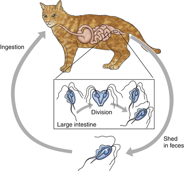
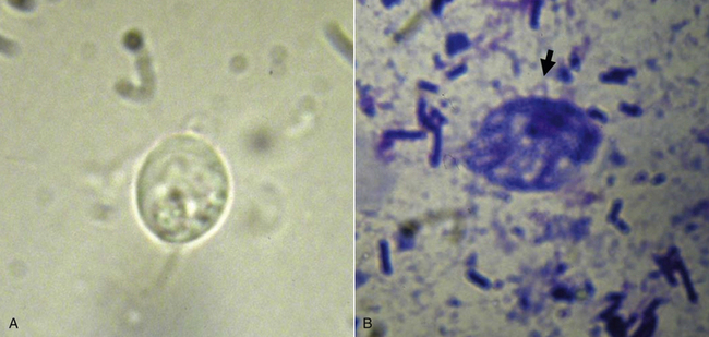
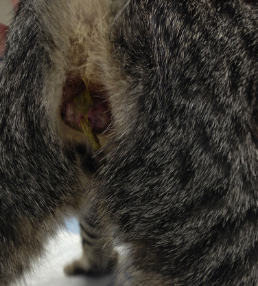
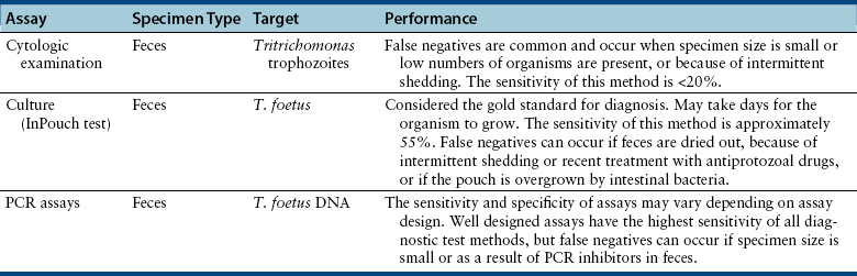
Chapter 80 Members of the order Trichomonadida are flagellates that reproduce by binary fission and do not form cysts (Figure 80-1).2,3 The trophozoite of Tritrichomonas foetus is 10 to 26 µm long and 3 to 5 µm wide and has three anterior flagella (Figure 80-2). In vertebrates, trichomonads are parasites of the gastrointestinal system, the reproductive system, and the upper portions of the respiratory tract. Tritrichomonas foetus infects the reproductive tract of both cows and bulls and can cause abortion and other reproductive abnormalities. The organism parasitizes the gastrointestinal system of the cat and can be associated with diarrhea. Genetic characterization of bovine and feline isolates of T. foetus has been performed, and minor differences exist.4–6 Feline isolates of T. foetus remained in the reproductive tract of heifers but did not cause the same pathologic changes as a bovine isolate in one study.7 Similarly, bovine isolates of T. foetus may not cause diarrhea in cats as successfully as feline isolates.8 FIGURE 80-1 Life cycle of Tritrichomonas foetus. The organism has a direct life cycle, and the parasite replicates by binary fission. FIGURE 80-2 A, Tritrichomonas foetus trophozoite seen on a saline wet mount of a fecal specimen. The organism can be recognized by its progressive forward, rolling motility. B, Diff Quik stain. The undulating membrane can be clearly visualized coursing along the lateral aspect of the trophozoite (arrow). Cats that harbor T. foetus infection have now been documented in multiple countries.9–27 Cats in crowded environments and purebred catteries are commonly colonized; the organism is uncommon in feral cats. In one of the first large prevalence studies in the United States, 31% of 117 cats owned by 89 different breeders at an international cat show were colonized.14 In the United Kingdom, 16 (14.4%) of 111 diarrhea specimens were positive for T. foetus by PCR assay.15 T. foetus–associated diarrhea is most common in kittens and young cats, but adult cats can also be affected.14,15 Catteries with a recent history of refractory diarrhea, adult cats with Isospora spp. infections, and cats living in a facility with a small number of square feet per cat were more likely to be infected with T. foetus in some studies.14,15 Other co-infections, such as those with Giardia spp. and Cryptosporidium felis, may also increase the potential for disease. Tritrichomonas foetus is an obligate parasite that depends on endogenous bacteria and host secretion for nutrients. However, the organism can survive for short periods of time in contaminated water, urine, feces, and cat litter.28,29 In one study, the organism was shown to survive 30 minutes on dry cat food, several hours in moist cat food, and hours to days in cat feces.29 The organism also survived transit through the gastrointestinal tract of two types of slugs.30 In experimentally infected cats, the location of T. foetus is restricted to the ileum, cecum, and colon.31,32 Fluctuations in the colonic microflora or other factors are necessary to produce the clinical signs of T. foetus infection. The organism can persist in cats without diarrhea, and diarrhea can worsen without a change in the organisms present, suggesting that the underlying mechanisms that lead to diarrhea are multifactorial.31 The organism adheres to the intestinal epithelium and incites a lymphoplasmacytic and neutrophilic inflammatory response.33 An increased number of T. foetus organisms and increased severity of diarrhea was associated with co-infection with Cryptosporidium spp. in one study.31 However, co-infections are not always associated with the presence of T. foetus–associated diarrhea.23 There is little information available as to whether immunosuppression predisposes to chronic infection with T. foetus. Administration of prednisolone did not change the fecal consistency or the frequency of positive results on direct fecal exams in one study.31 The clinical signs of trichomoniasis are variable and range from subclinical infection to chronic intractable diarrhea. The signs often are intermittent and can resolve with antimicrobial drug treatment, only to recur after treatment is discontinued. The diarrhea is mainly a large bowel diarrhea characterized by increased frequency of defecation and passage of semiformed to liquid, often foul-smelling feces that sometimes are associated with fresh blood and mucus. When the diarrhea is severe, the anus may become edematous and fecal incontinence may develop (Figure 80-3). Although diarrhea may persist, most affected cats maintain a good appetite and body condition score. FIGURE 80-3 Anal swelling and evidence of fecal incontinence in a kitten with suspected Tritrichomonas foetus infection. Tritrichomonas foetus was detected in the uterine contents of a cat with endometritis and pyometra.32 However, the organism was not associated with infection or disease of the reproductive tract in a separate study of cats with T. foetus infection that were housed in catteries.34 A direct fecal smear should be performed for all cats with large bowel diarrhea to evaluate for the presence of T. foetus trophozoites (Table 80-1), using the technique described for Giardia (see Chapter 79). Tritrichomonas foetus is similar in size to Giardia but can be differentiated by the presence of an undulating membrane, a rapid forward motion, the lack of a concave surface, and a single nucleus. Video clips that demonstrate the classic motility can be found online (http://www.cvm.ncsu.edu/docs/personnel/gookin_jody.html). The trophozoite and its undulating membrane may also be identified on fecal smears stained with Diff Quik stain. The sensitivity of a direct smear using specimens from naturally infected cats was only 14% in one study, so false negatives are common with this method.31 The sensitivity can be improved by analyzing multiple fecal smears. If T. foetus infection is still suspected after the initial work-up, culture can be performed using a commercially available culture system (InPouch TF, BioMed Diagnostics, White City, OR). In this system, a tiny amount of feces is inoculated into the pouch, which can be incubated in the clinic at room temperature and examined daily by microscopy for evidence of protozoal growth. The medium used does not support the growth of Giardia spp. or Pentatrichomonas hominis, and positive test results suggest infection by T. foetus.35 This assay is considered the gold standard for diagnosis of infection. However, it takes days for positive test results to develop in some cases. Thus, PCR assays are also used by clinicians to document current infection. Several PCR-based assays that are specific for T. foetus are also commercially available, and these assays are recommended for assessment of fecal specimens that were found negative by microscopy and fecal culture and to confirm that microscopically observed or cultivated organisms are T. foetus.36 PCR assays are available that are specific for T. foetus and do not amplify Giardia or P. hominis DNA.37 The DNA of P. hominis has been amplified from the feces of cats co-infected with T. foetus, but whether this potentiates T. foetus–associated clinical disease is currently unknown.37
Trichomoniasis
Etiologic Agent and Epidemiology
Clinical Features
Diagnosis
Microbiologic Testing
< div class='tao-gold-member'>
![]()
Stay updated, free articles. Join our Telegram channel

Full access? Get Clinical Tree


Trichomoniasis
Only gold members can continue reading. Log In or Register to continue
