The Respiratory System

CHAPTER 34 The Respiratory System


Diseases of the respiratory system are common reasons for presentation of neonatal and immature dogs and cats. The respiratory system represents a large percentage of the total surface area of the body exposed to the external environment. With extensive mucosal surfaces in the nasal passage and trachea, as well as the very large surface area of the respiratory bronchioles and alveoli, the respiratory system is a frequent site of entry for the major viral and bacterial pathogens likely to affect puppies and kittens. Respiratory diseases in the puppy and kitten can rapidly proceed to severe, life-threatening disorders if they are not recognized early.


Normal Respiratory System Development


The embryology of the respiratory system, including the nasal passages, larynx, trachea, bronchi, and alveolar tissue of the lungs, is highly complex. The primordial respiratory system arises as an outpouching of the embryonic foregut. This outpouching subsequently develops via elongation into the respiratory diverticulum. The respiratory diverticulum remains contiguous with the embryonic foregut during development, with the connection forming the pharynx. As the embryo develops, the respiratory diverticulum elongates and bifurcates at the blind end of the pouch, forming bronchial buds, the primordia of the lungs. The trachea subsequently develops from the section of the respiratory diverticulum cranial to the bronchial buds. The bronchial buds undergo extensive bifurcation to form the bronchial tree. Each bronchial bud bifurcates between 14 and 18 times, eventually resulting in the extensive system of terminal bronchioles. Each terminal bronchiole will then subdivide another two to three times to yield the respiratory bronchioles. Respiratory bronchioles represent a transitional zone between the air-conducting structures of the upper airways and the alveolar tissue, in which gas exchange takes place. At birth, the lungs are not fully developed. Alveolar growth and formation of additional respiratory bronchioles continue for some period after birth. In the puppy, almost all the alveolar stage (the final stage of lung development) occurs in the initial postnatal period.


During fetal life the lungs are not inflated and are not functional from a respiratory perspective. The fetal lung tissue is filled with fluid secreted by the pulmonary epithelial cells and mucosal glands. The presence of a normal quantity of this fluid within the developing lung is an important stimulus for ongoing expansion of the alveolar tissue. Insufficient production of this fluid is associated with pulmonary hypoplasia. During the later stages of fetal life, respiratory muscle activity commences. The fetus “breathes” fluid secretions from the pulmonary epithelium and a small amount of amniotic fluid. This breathing action prepares the respiratory muscles for activity in the postnatal period. In the last stages of fetal development, the pulmonary epithelium commences the synthesis of surfactant. This fluid forms a phospholipid layer over the alveolar surface, dramatically reducing the surface tension of the alveoli. Adequate surfactant formation is critical to the early stages of postnatal life. Inadequate surfactant production may be associated with acute respiratory distress in the newborn and in fading puppy or kitten syndromes.


In some scenarios when assisted mating and elective cesarean section delivery are planned, there may be uncertainty regarding adequate development of surfactant. Inadequate surfactant in the neonate will complicate peripartum management. While the puppies are still fetal and before delivery, the presence of surfactant can be indirectly documented using the “foam stability test.” This technique was originally used in human obstetric medicine as a rapid test to determine the timing of a cesarean section. One milliliter of amniotic fluid is collected (via ultrasound-guided centesis or laparotomy) and is mixed with 1 ml of 100% ethanol in a glass tube. The solution is then vigorously shaken for 15 seconds. If surfactant is present, a ring of bubbles will form at the fluid-air interface and remain for at least 15 minutes (Figure 34-1). In human obstetrics, this method was used as a surgery room test to indicate fetal readiness for delivery. Results of this test in elective cesarean section delivery of canine fetuses have indicated that adequate surfactant production is not present until 62 days past the luteinizing hormone (LH) surge for female canine fetuses and 63 days past the LH surge for male canine fetuses. This sex difference is consistent with observations in human obstetrics, as female fetuses begin surfactant production earlier than males.


Clinical Examination of the Respiratory System


It is important to remember that some aspects of respiratory system development continue to occur in the postnatal period. Thus physical examination findings of neonatal puppies and kittens are expected to differ from older puppies and kittens and the young adult animal. Typically puppies and kittens have more rapid breathing rates and shallower respiratory excursions than adults. The cardinal sign of respiratory disease is dyspnea or respiratory distress. Respiratory distress may manifest as increased respiratory rate, increased respiratory effort, decreased exercise tolerance, increased respiratory sounds, or acute collapse. The initial physical examination of an animal presented in respiratory distress should be brisk, thorough enough to provide initial diagnostic information, and carried out in a manner that causes minimal distress to the patient. In some circumstances, it may be appropriate to conduct the initial examination and triage without the animal’s owner present in the room to reduce the stress on both the patient and the owner.


Panting in dogs is a normal response to increased heat load and is not normally considered pathological. In the kitten or young cat, however, panting is almost always a sign of significant respiratory distress. Open-mouth breathing in both puppies and kittens is considered a strong indicator of respiratory distress. Animals presented in severe respiratory distress should receive supplemental oxygen therapy. This may be necessary before detailed examination can take place. Supply of supplemental oxygen using an oxygen cage is usually less stressful than via a mask and is more effective than flow-by oxygen delivery. If an oxygen cage is not available, however, all efforts should be made to supply supplemental oxygen in a minimally stressful manner. Some patients with dyspnea are panicked and struggle excessively. In these patients, sedation may be helpful and, in some cases, lifesaving.


Respiratory Patterns


Definition of the phase of the respiratory cycle showing the greatest alteration in respiratory effort can be helpful in defining an initial list of differential diagnoses and formulating a diagnostic plan. The general characteristics of the two main pathological respiratory patterns, obstructive and restrictive, are summarized in Table 34-1. Respiratory diseases that lead to restriction of the airway diameter typically present with an obstructive breathing pattern. In a patient presenting with a typical obstructive breathing pattern, respiratory rate is mildly to moderately increased, and the depth of respiratory excursions is usually increased. Obstructive breathing patterns may be seen with conditions affecting either the extrathoracic or intrathoracic airways. With obstructive lesions in the upper airways, inhalation tends to cause worsening obstruction as a result of decreasing airway diameter. Therefore patients with upper airway obstruction tend to show increased respiratory effort on inhalation. The reverse applies in lower airway obstructive diseases, as the diseased airways are more likely to collapse during exhalation, leading to increased expiratory effort. Respiratory diseases that limit the ability of the lungs and airways to expand lead to a restrictive breathing pattern. Inability to expand the lung parenchyma leads to a reduction in respiratory excursions. Patients presenting with restrictive breathing patterns show a shallow depth of respiration and increased respiratory rate in an attempt to maintain adequate ventilation. Restrictive breathing patterns may be seen with diseases affecting either the lung parenchyma or the pleural space. In particular, space-occupying lesions of the pleural cavity (e.g., pleural effusion, diaphragmatic hernia, pneumothorax, and pleuroperitoneal or peritoneopericardial hernias) may severely restrict the ability of the lungs to expand during inflation.


TABLE 34-1 Characteristics of obstructive and restrictive breathing patterns


Pattern Obstructive Restrictive
Respiratory depth Increased Shallow
Respiratory rate Normal to mild increase Markedly increased
Inspiratory dyspnea Extrathoracic airways Not typically distinguished
Expiratory dyspnea Intrathoracic airways Not typically distinguished

Thoracic auscultation is an integral part of the physical examination of animals with respiratory disease. The thorax of the neonate and very young puppy and kitten is much smaller than in adults. Auscultation with specific neonatal and pediatric stethoscopes is recommended to increase accuracy when attempting to localize respiratory sounds. The neonatal puppy has a normal respiratory rate at rest between 25 and 35 breaths/min for the first 2 weeks of life. Between 3 and 4 weeks of age, the normal respiratory rate decreases to between 15 and 25 breaths/min. After 4 weeks, most puppies show respiratory rates appropriate for adult dogs, including the onset of panting for thermoregulation. In theory, animals with upper airway obstructive disease have louder respiratory sounds on inhalation. Lower airway obstructive diseases (e.g., feline asthma) will tend to have louder respiratory noise on exhalation. In practice, it is often difficult to localize respiratory pathology with this degree of certainty. Even when signs of the respiratory pathology are localized to one area, this does not rule out the presence of disease in other regions. Animals with restrictive pulmonary diseases tend to have an overall reduction in the volume of breath sounds. Pleural effusions tend to settle to the ventral pleural cavity; therefore lung sounds may be muffled ventrally and prominent dorsally. In patients with pneumothorax, on the other hand, the lungs may settle ventrally, leading to muffled lung sounds dorsally. Although the lung sounds overall are decreased with restrictive diseases, auscultation directly over the partially collapsed lung tissue may reveal harsh respiratory sounds and pleural friction rubs. Common differential diagnoses of obstructive and restrictive breathing patterns are summarized in Box 34-1.


Congenital Anomalies of the Respiratory System


Brachycephalic Airway Syndrome


The brachycephalic airway syndrome is a common cause of respiratory distress in affected brachycephalic breeds. This condition is frequently identified in English Bulldogs, French Bulldogs, Pugs, Boston Terriers, Shih-Tzus, and Boxers. Although this condition is generally held to be heritable, the mode of inheritance has not been identified in any affected breed. Brachycephalic airway syndrome is much less commonly observed in brachycephalic cat breeds, but feline cases have been reported. Affected animals will display increased inspiratory effort and disordered respiration during sleep. Clinical signs are common from an early age in affected breeds. Essentially all British Bulldog puppies show abnormal respiration during sleep from the age of 2 weeks onward.


Primary lesions of brachycephalic airway syndrome are stenotic nares and soft palate elongation. Eversion of the laryngeal saccules is a common secondary condition. Tracheal hypoplasia, tracheal collapse, and laryngeal collapse, if present, are associated with more severe presentations. Development of the brachycephalic airway syndrome is related to the anatomical narrowing of the nasal passages and upper airways common to these breeds. Prolonged exposure of the airways to marked negative intraairway pressure leads to dynamic collapse of these airways, further decreasing the airway lumen and requiring greater respiratory effort. The soft palate further elongates, while the soft tissue of the pharynx swells and becomes redundant. Increased turbulence of airflow in the pharynx and upper airways acts as an inflammatory stimulus, exacerbating the soft tissue swelling. Soft tissue swelling and turbulent airflow are responsible for the characteristic marked stridor exhibited by these breeds.


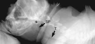
A diagnosis of brachycephalic airway syndrome should be suspected in any brachycephalic animal presented for respiratory distress. Diagnosis of the component disorders of this syndrome typically requires a combination of clinical examination, upper airway examination via laryngoscopy, and diagnostic imaging. Stenotic nares are readily visible during physical examination. Elongation of the soft palate can be documented via visual examination of the distal pharynx or radiographic examination of the upper airways (Figure 34-2). The normal soft palate should just overlap the epiglottis, whereas in brachycephalic airway syndrome the soft palate extends more than 3 mm caudal to the tip of the epiglottis and may completely occlude the epiglottic opening. In symptomatic individuals, early resection of the elongated soft palate is associated with a better long-term prognosis. Although the technique for surgical resection of an elongated palate is relatively simple, these patients represent a significant anesthetic risk and must be managed aggressively in the perioperative period to avoid swelling of the surgical site and acute upper airway obstruction. Placement of a temporary tracheostomy may be required in some patients.


Stay updated, free articles. Join our Telegram channel

Sep 11, 2016 | Posted by in SMALL ANIMAL | Comments Off on The Respiratory System

Full access? Get Clinical Tree

Get Clinical Tree app for offline access