Spleen removal is usually best performed via a laparotomy performed after the removal of the 17th rib. A 20-cm-long incision should be made over the 17th rib through the skin, subcutaneous tissues, and musculature. The periosteum should be sharply incised and reflected circumferentially from the rib using a periosteal elevator. The rib can then be luxated at the costochondral junction using a rib splitter and can then be reflected dorsally and transected at the most dorsal aspect of the abdominal incision using Gigli wire and then removed. This will allow entrance to the abdomen after extension of the incision through the axial periosteum and peritoneum. Gas insufflation should be discontinued at this point; however, the laparoscope and vessel-sealing device can still be used effectively to aid in hemostasis of the splenic artery and vein coursing into the hilus of the spleen. Hemostasis of this vasculature is critical and may be also accomplished with the use of extracorporeal knots added to the sealing effect of the LigaSure device. An enlarged spleen transection of the nephrosplenic ligament, viewed through the laparotomy incision, will facilitate access to the splenic hilus for improved sealing and ligation of the splenic artery and vein. Once these vessels are ligated, the spleen can be carefully removed from the abdomen. This procedure should be done with care since enlarged and friable spleens can fracture or rupture during manipulation (Roy et al. 2000; Ortved et al. 2008).
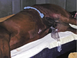
After removal of the spleen and verification of hemostasis at all transected vasculatures, routine closure of the laparotomy wound and laparoscopy portals can commence. Closure of the laparotomy wound should incorporate closure of the transverse abdominis and the internal abdominal oblique muscle, then the external abdominal oblique muscle, then the skin. Support and protection of the wound for recovery from anesthesia and immediate postoperative period should involve placement of a stent bandage to minimize swelling and to reduce subcutaneous dead space formation (Figure 32.2).
Figure 32.2 Horse positioned in right lateral recumbency following laparoscopic-assisted splenectomy. Note stent bandage applied over the left flank laparotomy.
Postoperative Management
Horses that have undergone splenectomy may require a blood transfusion depending on the estimated blood lost due to either the initial insult necessitating splenectomy or intraoperative hemorrhage (Roy et al. 2000; Garcia-Seeber et al. 2008; Muurlink et al. 2008; Ortved et al. 2008). Provisions for a blood transfusion should therefore be made both intra- and postoperatively.
Horses that have undergone laparoscopic splenectomy may demonstrate mild signs of abdominal pain for the first few days (Ortved et al. 2008). In addition to routine administration of non-steroidal anti-inflammatory drugs, some horses may benefit from additional analgesia such as epidurally administered morphine and/or a constant rate infusion of analgesic medications such as lidocaine, butorphanol, ketamine, or a combination thereof.
Broad-spectrum postoperative antimicrobial therapy should be administered due to the potential risks of sepsis, having removed an organ important for immune function. In adult horses, intravenous antimicrobials have been administered for 1 week postoperatively (Ortved et al. 2008), and in foals, antimicrobials have been administered for 1.0–3.5 weeks postoperatively (Garcia-Seeber et al. 2008; Muurlink et al. 2008).
Stay updated, free articles. Join our Telegram channel

Full access? Get Clinical Tree


