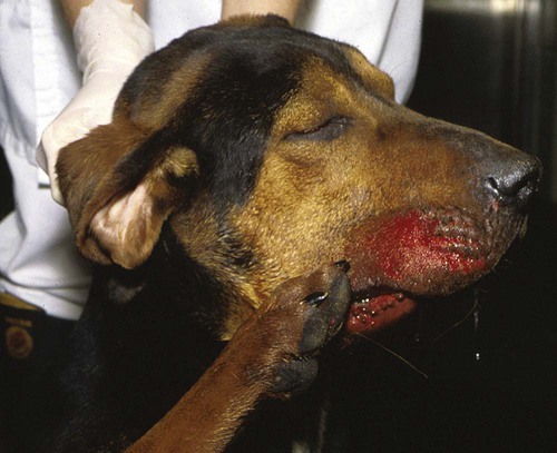
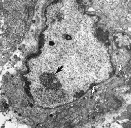
Pseudorabies virus (PRV), also known as porcine herpesvirus-1, is an enveloped DNA virus and an α-herpesvirus. As with other herpesviruses, PRV can cause latent infection, with viral DNA being incorporated into the host cell genome. The virus is relatively resistant to environmental factors and can survive outside the host for several months under favorable climatic conditions. Survival of PRV depends on temperature (10 days at 37° C, 40 days at 25° C) and pH (optimum, 7), and it is quickly inactivated by drying and exposure to ultraviolet light.6 The molecular biology of PRV has been extensively studied because PRV is probably one of the most used viruses in experimental neuroscience.38 PRV infection (known as Aujeszky’s disease, mad itch, and infectious bulbar paralysis) occurs in most countries of the world with the exception of Australia and has been responsible for massive economic losses to the swine industry. In the past, clinical disease outbreaks have been reported in several European, South American, and Asian countries. Restricted endemic areas are in the United States, Mexico, Brazil, and China.55 However, a PRV surveillance scheme is lacking in many countries. Several European countries in which the disease occurred previously are now considered to be PRV-free. The latter is undoubtedly due to systematic vaccination of the pig population. Although many mammalian species are susceptible to infection with PRV, it is predominantly a problem in pigs, the main reservoir of the virus. However, cattle, fur-bearing animals, goats, sheep, dogs, and cats are sporadically affected.18,27,27 It does not appear to affect humans; most reports have been circumstantial and not documented. Infection frequently is subclinical in pigs because they have become well adapted to the virus. The disease is spread by commercial movement of infected pigs or contaminated pork products. Venereal transmission occurs when infected boars shed PRV in semen. Although wild animals such as raccoons, panthers,12 and rats may act as transient reservoirs, they are not important in maintaining the disease in nature. Their role is limited to temporary local spread of virus within enzootic areas. However, in several countries such as Switzerland, Spain, and the United States, PRV infection is endemic in wild boars, creating a reservoir for the virus.24,51,51 PRV infection in dogs and cats is thought to occur only in areas where the disease is enzootic in pigs. Such epidemiologic interaction among species has been documented by molecular biological studies on viral isolates.13 In fact, the occurrence of typical pseudorabies signs in pets can be the first indication that the disease is enzootic in the local pig population. Pets almost invariably are infected as a result of consuming contaminated raw pork. Dogs also have developed pseudorabies after having bitten infected pigs. Direct spread from dog to dog has not been shown to occur.37 However, one report described an outbreak in animals without direct contact with swine. Isolated strains of PRV showed considerable differences in DNA patterns to vaccine and isolated strains obtained from pigs in the same region.42 Only a few feline and canine cases have been reported in Poland, Italy, Austria, Germany, and the United States in recent years.3,16,23,42,44 The disease seems to be very rare in the eastern part of Europe where PRV infection used to be widespread,32,45 as well as in north America.26 One canine case in Portugal in 2003 and 12 cases in Bulgaria in 2004 have been recorded.55 Naturally acquired infection in dogs and cats occurs after ingestion of the virus, although a similar sequence of events occurs after parenteral inoculation of virus. PRV enters the nerve endings at the inoculation site and travels in retrograde fashion via the axoplasm of the nerve fibers to the brain. The incubation time in dogs and cats, regardless of inoculation sites, is 3 to 6 days. Experimental studies in orally infected cats have shown that PRV replicates in the tonsils and travels from the oral mucosa via the sensory branches of the 9th and 10th cranial nerves to the nucleus tractus solitarius and area postrema in the medulla oblongata.17 The 5th cranial nerve has been less frequently involved. Experimental infections in rats have shown that PRV spreads in a highly specific manner through synaptic connections.5,9 Viral–cellular interactions have been studied in great detail in experimental models.38 The initial sign often noted by the owner is a change in behavior, such as inactivity, lethargy, and indifference, although some animals become aggressive or restless. Dyspnea, diarrhea, and vomiting are occasionally seen. The dyspnea is often caused by severe pulmonary edema. Body temperature may be normal or abnormal, and hypersalivation is common. However, the most characteristic sign is intense pruritus, which usually occurs in the head region and rarely occurs in other areas such as the neck and shoulders. The animals violently scratch their faces and ears and rub their heads against the floor or walls. One side of the head and neck may become swollen. Self-mutilation results in erythema, excoriation, and ulceration of the skin and underlying tissues (Fig. 21-1). The scratching becomes increasingly frantic and may end in generalized convulsions. Most of the other neurologic signs that are observed in PRV infection refer to lesions in the lower brainstem and consist of one or several deficits in cranial nerve function. These deficits are usually unilateral and include anisocoria, mydriasis, lack of direct or consensual pupillary light reflexes, trismus, paresis and paralysis of the facial muscles, head tilt, inability to swallow, and vocal changes. Anisocoria and a hoarse voice are considered to be highly consistent signs in the cat.33 Less commonly observed neurologic signs include behavioral abnormalities such as aggressiveness, generalized hyperesthesia, head pressing, and generalized convulsions. The latter often occur as sequelae to frantic scratching. Paresis and paralysis of the limbs are sometimes noted shortly before death. Although less common, in some cats death has been preceded predominantly by acute gastrointestinal signs.52 Arrhythmias may manifest by an irregular pulse and dropped beats as a result of myocarditis.47 An atypical course of the disease has also been observed in cats that died suddenly without developing neurologic signs.21 Pruritus has been absent in some cases of spontaneous PRV infection in dogs54 and in experimental oral infection in cats.14 Gastrointestinal signs have been the predominant feature of some infected dogs.11 Hematologic or biochemical abnormalities are not found in pseudorabies. Electrocardiographic findings may include cardiac arrhythmias.31,47 Cerebrospinal fluid may show increased protein concentration and mononuclear pleocytosis. This finding is strongly indicative of viral encephalitis but is not specific for pseudorabies. Virus neutralization, immunodiffusion, and enzyme-linked immunosorbent assay methods commonly are used to detect serum antibody to PRV in pigs. Serologic studies have been valuable in determining the prevalence of disease in pig populations. Virus-neutralizing antibodies have not been found in sera from dogs tested during an outbreak of PRV infection.22 Diagnostic methods include fluorescent antibody examination for viral antigen in smears or frozen sections of various tissues. The brain and tonsils are the tissues of choice in such studies. Various polymerase chain reaction (PCR) techniques for different viral genes are also effective to detect PRV.25,30,30 PCR has been used successfully to detect the virus in cats and dogs.19,42 PCR is the method of choice to detect the presence of PRV in the porcine population.41,43 Virus can be isolated in tissue culture from lung and spleen and especially from brain and tonsils of animals with pseudorabies (Fig. 21-2). Although many cell lines have been used, most laboratories use pig kidney epithelial cells. A definite cytopathic effect consisting of syncytial formation is visible after 12 to 24 hours (Fig. 21-3). Virus isolation is not always easy in dogs, even in well-substantiated cases.2 Pharyngeal washings, tonsillar swabs, and saliva are unsuitable for viral isolation in dogs.22
Pseudorabies
Etiology
Epidemiology
Pathogenesis
Clinical Findings
Diagnosis
![]()
Stay updated, free articles. Join our Telegram channel

Full access? Get Clinical Tree


Veterian Key
Fastest Veterinary Medicine Insight Engine