Preprocedure Considerations


Preprocedure Considerations


This chapter covers important topics common to both chemical restraint and anesthesia. Understanding the information contained in this chapter will improve safety and efficacy when employing the techniques presented in subsequent chapters.


Preprocedure Considerations


Physical Examination


In the field setting a brief physical examination should be preformed, when patient cooperation allows, prior to using a potent chemical restraint technique or anesthetizing a camelid patient. This should include an overall assessment of the patient’s health status, auscultation of the cardiopulmonary systems, and evaluation of locomotor function. This helps determine whether the patient is a suitable candidate and perhaps reduce liability should complications occur.


In the hospital setting, packed cell volume (PCV) and total protein (TP) values should be determined in all patients that can be reasonably sampled prior to anesthesia. A complete blood cell count (CBC) and fibrinogen level determination improve the preanesthetic screening process, but for routine procedures in apparently healthy patients, economic and time constraints may limit the laboratory work performed. In all other patients a CBC and fibrinogen level determination should be performed. Additional laboratory work (serum chemistries etc.) should be based on patient status.


Site Selection and Facility Requirements


A flat, even surface that provides good footing should be selected. The surface should be soft to reduce risks of injury during induction and recovery. In the field setting, an open grassy area is generally ideal. In the hospital setting, an area with a resilient floor such as hoof matting should be used, if available. A stall deeply bedded with shavings may be used, although the confined space may interfere with the procedure and increase risk to personnel when larger camelids are involved. The site selected should be free of hazards in all directions for a reasonable distance. Larger camelids are more difficult to control physically and require a somewhat larger “safety zone.”


Camelids tend to be patient during recovery from anesthesia. They typically do not attempt to stand until they are awake and functional. Good footing is the primary requirement for achieving a good recovery. Proximity to water, electricity, and vehicle (containing emergency supplies) are also factors to consider when selecting a field site. A calm environment reduces patient anxiety and is always desirable.


A small animal surgery table may be used for alpacas and smaller llamas. A foal or small ruminant surgery table or a heavy-duty gurney will be required for larger llamas. A foam pad should be used to cushion the table surface. A shallow trough shaped pad (4-inch base thickness) makes dorsal recumbency easier to maintain and, in many cases, may be used for patients in lateral recumbency. A large animal surgery table is required for larger camels. Llamas and alpacas are typically transferred from the floor to the surgery surface and back manually. It is important to ensure that adequate personnel are available for intended transfers. A 1- to 2-ton overhead hoist may be used to move larger camelid patients into a better working position on the floor (preferably on a foam pad) or onto a surgery table. If the ceiling will not support an overhead mounted hoist without extensive modification, a lateral pull using a wall-mounted winch system may be used to reposition larger patients. Multiple mounting points and a portable winch allow patients to be moved to various areas of the operating theater. Two sets of hobbles connected by rings to the hook of the hoist or winch are used to move the patient. Hobbles are traditionally placed on the pastern but may be positioned above the fetlock, if required, to ensure a secure grip on the legs. Hobbles may be fashioned by splicing loops at each end of short segments of 1-inch cotton rope. The body of the rope segment is then fed through each loop to create the sliding component of the hobble. The distance between the loops needs to be sufficient to make placing and removing the hobbles easy, but excessive length reduces the height to which the patient can be lifted using an overhead hoist. Canvas strap hobbles (Shanks Veterinary Equipment, Inc., Milledgeville, IL) are available commercially. The wider, stiffer, canvas strap hobbles require more attention to attain a snug and secure fit. Good facility design makes managing larger patients safer and requires fewer personnel.1


Patient Positioning


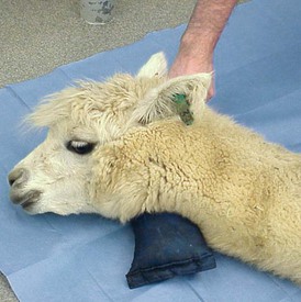
Camelids continue to salivate when sedated or anesthetized. The degree of protective laryngeal and eye reflexes retained will depend on the chemical restraint or anesthesia technique used and the drug doses selected. Camelids tend to lie down with heavier levels of sedation. Camelid patients that remain standing or are able to maintain sternal recumbency when chemically restrained typically clear saliva effectively. Patients in lateral or dorsal recumbency should be positioned such that saliva runs out of the mouth rather than pooling back near the larynx. In patients in lateral recumbency, this may be accomplished simply by placing a pad under the head–neck junction such that the opening of the mouth is below the level of the larynx (Figure 44-1). A mild downward tilt is generally all that is needed, as use of an overly large pad may impair venous return from the head, resulting in edema formation.


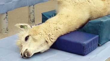
Protecting the airway becomes much more challenging when the patient is placed in dorsal recumbency. Camelid patients placed in dorsal recumbency will typically be under the influence of a potent chemical restraint protocol or anesthetized. The body should be elevated to produce a slightly dependent head position. The neck should be gently twisted so that the head rests laterally on the surgery table or floor (Figure 44-2). A shallow trough shaped pad (4-inch base thickness) provides the proper height and makes dorsal recumbency easier to maintain. This positioning places the mouth opening below the level of the larynx to facilitate saliva egress. The long neck of camelids makes this fairly easy to accomplish. A small pad placed under the head–neck junction may be needed to achieve the proper degree of head tilt. The head must be supported, rather the left hanging, to prevent excessive tension on vital neck structures and reduce the extent of edema formation. Placing the head in a dependent position increases hydrostatic pressures within the blood vessels of the head, which, over time, results in edema formation of the nasal, pharyngeal, and laryngeal tissues, as well as the eyelids. Minimizing the degree of dependence of the head relative to the body to only what is necessary to provide saliva egress may help to reduce the degree of edema formation.


The head and neck of a recumbent patient should be extended to minimize impingement of pharyngeal tissue into the airway in a nonintubated patient and prevent kinking of the endotracheal (ET) tube in an intubated patient. Nasal edema is probably the most commonly recognized complication of anesthesia in camelid patients. Camelids with severe nasal edema are able to mouth-breathe but appear stressed when this occurs. Patients that are chemically or medically obtunded may not be able to effectively compensate. It is important to monitor nonintubated patients under the influence of potent chemical restraint or injectable anesthetic protocols and patients recently extubated for signs of respiratory compromise. One must be prepared to respond with ET intubation, if required. In patients exhibiting mild-to-moderate difficulty breathing during recovery, elevating and gently extending the head and neck in a manner that still facilitates saliva egress helps alleviate edema and maintain a patent oral airway. Airway edema generally resolves relatively quickly once the patient is able to keep its head up. Nasopharyngeal intubation, instillation of decongestant products, or both are techniques that have been used to counter the impact of severe nasal edema. Respiratory compromise resulting from laryngeal edema is less common but may be life threatening. Patients exhibiting severe respiratory distress should be endotracheally intubated. Additional information on patient positioning is provided in the “Oxygen Delivery, Muscle and Nerve Protection” section of this chapter.


Endotracheal Intubation


The camelid patient should be intubated when the head cannot be positioned to facilitate salvia egress or the procedure could result in blood or other material entering the airway. Intubation is also required to deliver inhalant anesthetics (saliva production precludes safe mask delivery of oxygen or inhalants).


Alpacas and llamas are intubated by direct visualization, as in dogs and cats. Because of the small mouth opening and deep oral cavity in camelids, proper alignment is important to visualize the larynx (Figure 44-3). This is similar to looking through a long, narrow tube. A laryngoscope with an extra-long blade aids visualization of the larynx by allowing greater control of the base of the tongue (Wisconsin 11-inch, 14-inch, and 18-inch laryngoscope blades; Anesthesia Medical Specialties, Beaumont, CA) (Figure 44-4). The ET tube must be long enough to place its inflatable cuff below the level of the larynx in the trachea. Oral cavity depth of larger alpacas and llamas typically requires the use of longer 5- to 14-mm silicone “foal” ET tubes (Bivona Aire-Cuf Standard Silicone Endotracheal Tubes with Murphy Eye and Connector, Smiths Medicals). A selection of ET tube diameters should be available to ensure proper fit.
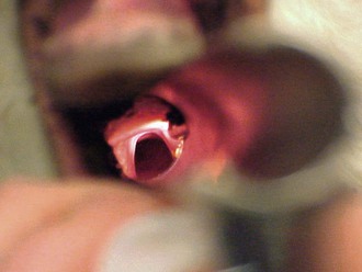
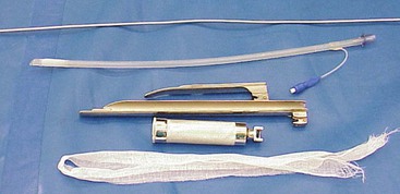


Keeping the patient in sternal recumbency with the head elevated reduces the risk of passive regurgitation during intubation. It is important to keep the patient’s mouth pointed downward until the intubation process is imminent to minimize pooling of saliva back around the larynx. An assistant straddles the patient’s back using his or her lower legs to hold the patient in sternal recumbency. The assistant extends the head and neck up toward the individual doing the intubation and holds the jaws apart. The assistant’s knees may be used to help control the patient’s head and neck. A reduced level of jaw tone and the absence of the chewing or lingual response to oral manipulation are used to determine when intubation is appropriate. Sliding a large camelid patient down a wall is safer for personnel involved and improves the odds of maintaining sternal recumbency for the intubation process. Additional information on preventing regurgitation is provided in the “Fasting before Anesthesia” section of this chapter.


A stylet (one eighth–inch aluminum rod for larger tube sizes and two guttural pouch cannulas glued end-to-end for smaller tube sizes) is used to facilitate intubation of camelids. The thin stylet does not obstruct the view of the larynx. The ends of the stylet should be smooth or rounded to minimize the risk of damaging the mucosal surfaces of the airway. The stylet is guided into the larynx first and the ET tube passed over it into the airway. The stylet must be long enough that it can be grasped above the ET tube as it is advanced down the length of the stylet and into the trachea. With practice, the ET tube can be positioned on the stylet and held in place, with the hand guiding the stylet into the airway, making the process less cumbersome. In large llamas, the size of the oral cavity may be large enough to allow visualization of the larynx while the ET tube is guided into the airway. The stylet is used to stiffen the ET tube. Allowing the stylet to protrude slightly from the end of the ET tube makes placement in the airway easier.


Because of the depth of the oral cavity, the presence of glottal folds, and the size of the ET tube required, larger camels are intubated by manually guiding the tube into the airway, as in adult cattle. The anesthetist carries the ET tube into the mouth with one hand and then uses his or her fingers to guide the tube between the arytenoids as the other hand advances the tube. A speculum is required for this technique. The arm or hand size of the individual performing the intubation is the limiting factor in determining the appropriate patient size for this approach. The oral cavity must be large enough to accommodate the operator’s arm and the ET tube. The lower limit for this technique is generally around 300 to 350 kilograms (kg), unless the operator has an exceptionally small arm and hand. A somewhat undersized ET tube may provide additional room for the operator’s arm in marginally sized patients. The operator should wash the arm thoroughly afterward because camelid saliva may irritate the skin of some individuals.


A flexible endoscope may be used to guide the ET tube into the larynx. The endoscope serves as both a stylet and a visualization device. This technique has proven useful when traditional methods cannot be used. The endoscope must be small enough to fit inside the ET tube.


Camelid patients that are actively spitting prior to anesthetic induction seem to be more prone to regurgitation compared with nonspitters. Feed material is often trapped in the mouth following induction, so extra care must be taken to ensure that it does enter the airway. The small diameter of the tube used in traditional medical suction devices may make removing the coarse feed material difficult at times. Often the fastest and most effective method for clearing the material is to scoop it out using the blade of the laryngoscope. Unfortunately, fasting does not seem to eliminate this rather unpleasant behavior.


Intravenous Catheters Are “Expensive” and Time Consuming: Are They Really Necessary?


A catheter is generally not required when using simple chemical restraint techniques. A catheter should be placed prior to anesthesia in camelid patients. The jugular vein is the most commonly used site for IV catheter placement. The thick fiber coat and neck skin of camelids, combined with poor patient cooperation, may make catheterization of the jugular vein challenging, and carotid sticks do occur. A 14-gauge 5.5-inch catheter is used in most camelid patients. An 18-gauge 2-inch catheter is sufficient for crias. The catheter should be secured to the neck with suture, bandage, or both. The catheter should always be checked before anesthetic induction to ensure that it is still functional.


The veins on the external surface of the ear of some camelids are relatively large and accessible. Ear veins can be used to deliver small-volume IV chemical restraint cocktails. Most patients require only modest head restraint when a 25-gauge needle and good technique are used. Ear veins may be used as an alternative site for venous catheter placement. An 18- gauge, 20-gauge, or 22-gauge catheter should be used, depending on the vessel size, and secured with a combination of super glue and tape. Because of the increased level of stimulation produced by the catheter stylet, camelid patients are typically less than cooperative to ear catheterization unless chemically or medically obtunded. The cephalic vein may also be used as a site for venous catheter placement, much as it is in small animal patients. The smaller catheters will not provide the flow rate of the 14-gauge catheter but work well otherwise.


Camelid owners are frequently concerned about the clipping of fiber, which makes venous catheterization somewhat more challenging. One approach practitioners may find useful is to administer a Ketamine Stun via the ear vein and then place an IV catheter once the patient is recumbent and cooperative. Double Drip or Ruminant Triple Drip may then be delivered to maintain injectable anesthesia or facilitate ET intubation for inhalation maintenance.

Stay updated, free articles. Join our Telegram channel

Mar 27, 2017 | Posted by in GENERAL | Comments Off on Preprocedure Considerations

Full access? Get Clinical Tree

Get Clinical Tree app for offline access