CHAPTER 72 Pregnancy Diagnosis in Goats
Caprine pregnancy can be diagnosed over a wide range of times during gestation. Demand for pregnancy diagnosis, however, varies with herd type. Owners may simply want to know if their goats are pregnant. Commercial milk producers may justify examination costs with costs of keeping open does and the need to maintain production. Intensively managed herds benefit from early diagnosis by maximizing the number of pregnancies during a defined period. Goats bred out of season have variable pregnancy rates and diagnosis allows planning for winter milk supplies. Pregnancy diagnosis also helps monitor the success of artificial insemination.
PITFALLS TO CLINICAL DIAGNOSIS
An accurate caprine pregnancy test must distinguish hydrometra from pregnancy. Hydrometra occurs commonly in goats and may cause false positive results. Annual herd incidence varied between 3% and 21% in one study, with considerable variation among herds and within herds during different years.1 The prevalence of hydrometra is perhaps the most important reason for pregnancy diagnosis in commercial dairy herds. The test must also be highly sensitive (few false negatives). Pregnant goats given prostaglandin usually abort. A false negative test in a doe given prostaglandin will become readily apparent.
Estrous Behavior and Physical Appearance
Goat owners frequently use clinical signs such as estrous behavior following breeding and abdominal contour to make a presumptive diagnosis of pregnancy. Failure to return to estrus after breeding during the ovulatory season suggests pregnancy, but does with hydrometra or pathologic ovarian conditions may also fail to exhibit estrus. Failure to return to estrus cannot be relied upon when does are bred late in the breeding season or during the anovulatory season. Pregnant does may show standing estrous behavior, further making this an unreliable sign. Goats with hydrometra may have large, pendulous abdomens and look pregnant. Goats with permanent stretching of the abdominal muscles (dropped stomach) have distended, pendulous abdomens and could be mistaken for pregnant.2
Abdominal Palpation and Ballottement
During the last half of pregnancy, the gravid uterus or fetus can sometimes be palpated through the abdominal wall. This is difficult in does that tense their abdominal muscles. Ballottement of the fetus low in the right flank, as performed in cattle, may be attempted in late pregnancy, but one is often left wondering if the “bump” felt was the ventral sac of the rumen or a fetus. Rectal-abdominal palpation with a rod has been done in goats but was too traumatic to be safely recommended.3
A bimanual palpation technique was recently described wherein the reproductive tract is examined per rectum by the index finger of one hand while the fingertips of the other hand are pushed upward into the ventral floor of the posterior abdomen.4 Pregnancy was diagnosed from day 28 onward with this method. At an early stage, hydrometra cannot be distinguished from pregnancy with this technique.
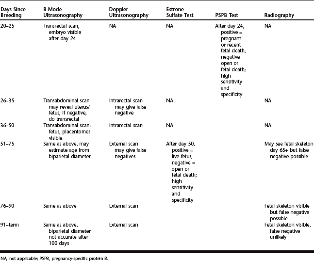
Recent technologic advances have replaced many of the previously described caprine pregnancy tests.5 Realtime ultrasonography and biologic tests are now the most sensitive and specific methods. Tests specific for pregnancy in the goat and their application relative to the day of breeding are summarized in Table 72-1.
ULTRASONOGRAPHY
A-Mode Ultrasonography
Ultrasound waves emitted by amplitude-depth units are reflected from tissue interfaces and converted to audible or visual signals. These units detect fluid-filled organs at a depth of 10 to 20 cm. Pregnancy diagnosis is based on finding fluid within the uterus, so hydrometra or pyometra will give false positive results. False negatives are possible during late pregnancy when the volume of uterine fluid relative to fetal size has diminished. Accuracy is affected by the prevalence of hydrometra in the herd. Lack of specificity for pregnancy makes A-mode ultrasonography unreliable for pregnancy diagnosis in goats.
Doppler Ultrasonography
Transabdominal scans are done by placing the transducer low on the right flank. Transrectal scans are done by inserting a specially designed rectal probe into the rectum and slowly rotating it in the area of the pelvic inlet and caudal abdomen. Fetal sounds include pounding of the fetal heart, sharp fetal movements, and the swishing sound of blood flow in the umbilical arteries.6,7 Maternal sounds include swishing blood flow in the middle uterine artery and rumbling intestinal movement.6,7
During the first half of gestation, transrectal Doppler was more accurate than the external approach.6 Transrectal scans can be attempted at 25 to 30 days after breeding, but false negative results are a problem.7 It is preferable to wait until day 35 to 40. Accuracy of transrectal Doppler was 94% to 100% for detecting pregnancy and 25% to 75% for detecting nonpregnancy in goats 55 or more days after breeding.3 During the last half of gestation, external Doppler was nearly 100% accurate for detecting pregnancy.6 A 2.25-MHz transducer is suggested for near-term pregnancies and a 5-MHz transducer for earlier pregnancies.6 B-mode ultrasonography has largely replaced Doppler for pregnancy diagnosis.
B-Mode Ultrasonography
B-mode ultrasound units produce a realtime, two-dimensional image on a screen, allowing direct visualization of the uterus, fetal fluids, fetus, fetal heartbeat, and placentomes. A 5-MHz linear-array transducer is the most versatile for reproductive work in livestock. It can be used externally or intrarectally over a wide range of gestational ages. Lower frequency linear-array, sector, or curvilinear probes are also suitable for external use in goats. B-mode scanning for pregnancy can be rapidly learned, and experienced examiners achieve an accuracy of 91% to 100%.8,9
Transrectal scanning is done by inserting the lubricated probe into the rectum and slowly rotating it from side to side. Transrectal scanning is usually reserved for diagnosis at less than 30 to 35 days after breeding. Some does object at first but most acquiesce in a short time. The anechoic urinary bladder serves as a good point of orientation and is recognized by its triangular neck as the probe is moved caudally. A poor quality image or no image is usually due to feces trapped under the probe, in which case it should be removed, wiped clean, and reinserted. Limited maneuverability of the probe allows effective imaging only of the pelvic canal and an area just cranial to the pelvic brim. Alternatively, a transvaginal approach using a cylindrical sectoral probe with a variable angle head can be used to scan the reproductive tract from the cranial vagina.10
Pregnancy can be detected transrectally in most does by about 20 days after breeding.9 The embryo is often visible by 24 days and is seen more consistently as gestation advances.9 The ideal time for transabdominal scanning is roughly from 40 to 75 days of gestation when the pregnant uterus lies against the right body wall. Before day 40, the probe may have to be placed higher in the inguinal region and aimed toward the pelvic brim. Diagnosis of nonpregnancy less than 30 days after breeding is probably best achieved by the transrectal approach. Transabdominal scanning during the third trimester may require moving the probe cranially along the ventral abdomen to image the gravid uterus that has descended well into the abdominal cavity.
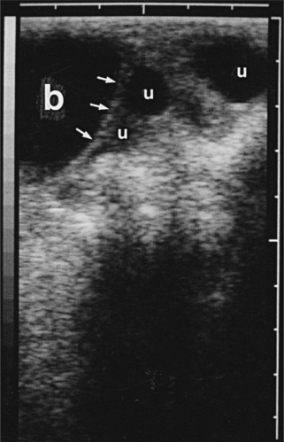
A positive diagnosis of pregnancy is made when the embryo, fetus, or placentomes are seen. A presumptive diagnosis of early pregnancy can be made when multiple fluid-filled sections of uterine lumen are seen cranial to the bladder (Fig. 72-1); however, hydrometra causes a false positive test. Hydrometra occurs commonly enough to advise cautious positive diagnoses until the embryo, fetus, or placentomes are seen.
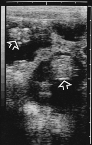
During the first trimester, the embryo appears as an echogenic mass surrounded by anechoic fluid within the uterine lumen (Fig. 72-2). Seeing fetal movements or the fetal heartbeat assures viability. During the second trimester, the fetal head, thorax, and limbs become more discernible. As pregnancy advances, only portions of the fetus such as the thorax or skull may be imaged at a time. As the ratio of uterine fluid to fetus decreases during late pregnancy, it may be difficult to distinguish the fetus from the rest of the dam’s abdomen. The fetal skull and beating heart are good points of orientation when scanning during late pregnancy.
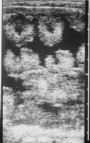
Placentomes are seen by 35 to 40 days as echogenic densities in the uterine wall. By 45 to 50 days, they are more easily seen as C-shaped densities with the concave surface directed toward the uterine lumen. As pregnancy advances, numerous placentomes appear as distinct donut-shaped and C-shaped echodensities surrounded by anechoic fluid when imaged in the longitudinal and cross-sectional planes, respectively (Fig. 72-3). During the second and third trimesters, these large placentomes are often seen immediately after the probe is placed on the skin and allow for a rapid diagnosis of pregnancy.
Stay updated, free articles. Join our Telegram channel

Full access? Get Clinical Tree


