Pathophysiology of heart failure

Chapter 5 Pathophysiology of heart failure


MECHANISMS OF HEART FAILURE


Heart failure is a clinical syndrome that can be caused by a variety of etiologies and mechanisms. The most common haemodynamic mechanism responsible for heart failure in horses is volume overload caused by valvular regurgitation or by a left to right shunt. However, myocardial systolic failure (reduced contractility) is also recognized in horses and may lead to heart failure. Less commonly heart failure is the result of impaired diastolic function as a result of inadequate cardiac filling in horses with pericardial disease.


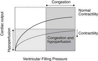
Regardless of the etiology or mechanism of heart failure, CHF is associated with two primary haemodynamic abnormalities: (1) increased ventricular filling pressures and (2) reduced cardiac output. Increased ventricular filling pressures are responsible for signs of congestion and oedema; whereas reduced cardiac output causes clinical signs of inadequate tissue perfusion. The haemodynamic abnormalities and resulting clinical signs that characterize heart failure are illustrated using ventricular function curves in Figure 5.1.


Horses with left-sided failure have increased left ventricular filling pressures and subsequently increased left atrial and pulmonary venous pressures leading to pulmonary oedema. Thus, clinical signs may include tachypnea, dyspnea, coughing, haemoptysis and the appearance of frothy white or blood-tinged fluid at the nostrils. Horses with right-sided CHF have increased right ventricular filling pressures and subsequently increased right atrial and systemic venous pressures leading to peripheral oedema. Subcutaneous oedema in the dependent areas such as the ventral abdomen, the prepuce and the distal extremities is the most common manifestation of increased right-sided filling pressures. Jugular venous distension and pulsation may also be present. In horses, an increase in left-sided filling pressure frequently stimulates pulmonary vasoconstriction and pulmonary vascular remodelling, changes that result in secondary right-sided heart failure. Thus, in some horses, systemic oedema and jugular distention may be the first clinical signs of chronic left-sided heart disease. Reduced cardiac output causes clinical signs referable to poor tissue perfusion, namely fatigue, weakness, exercise intolerance, cold extremities, prolonged capillary refill and hypothermia. With the exception of exercise intolerance and fatigue, these signs are not likely to become apparent until heart failure has become severe. Laboratory evidence of reduced cardiac output includes reduced venous oxygen tension in patients that are not hypoxaemic or anaemic as well as azotaemia and lactic acidosis if cardiac output is markedly decreased. (image AF, PC, VMD)

< div class='tao-gold-member'>

Stay updated, free articles. Join our Telegram channel

Jul 31, 2016 | Posted by in INTERNAL MEDICINE | Comments Off on Pathophysiology of heart failure

Full access? Get Clinical Tree

Get Clinical Tree app for offline access