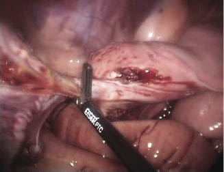
The ovary is grasped with the grasping forceps and the proper ligament of the ovary is transected with the coagulating/dividing device (Rodgerson et al. 2001; Lloyd et al. 2007; Smith & Mair 2008). The mesosalpinx and the oviduct may be also transected at this point or later. The ovary is now only attached by its blood supply dorsally. The coagulating/dividing device is then applied across the mesovarium and used to dissect the ovary free from its blood supply and attachments (Figure 29.2). The tension on the mesovarium is relaxed to check for inadequately sealed vessels frequently during the dissection. Once the ovary is freed from its attachments, the transected structures are inspected for hemorrhage. If hemorrhage precludes observation of the stump, a combined suction lavage cannula may be used to aspirate the blood.
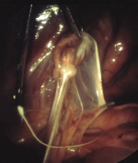
Other methods of vascular occlusion, including the Harmonic scalpel (Düsterdieck et al. 2003) and ultrasonic shears (Alldredge & Hendrickson 2004), have been reported and are effective. Ligating loops or Roeder knots and laser (Palmer 1993) or polyamide tie-wraps (Cokelaere et al. 2005) may be used around the ovarian pedicle after transection of the proper ligament and the mesosalpinx and oviduct (Figure 29.3). Endoscopic stapling and dividing devices are also used but are costly (Van Hoogmoed & Galuppo 2005).
Stay updated, free articles. Join our Telegram channel

Full access? Get Clinical Tree


