Osteochondrosis

CHAPTER 114 Osteochondrosis


Osteochondrosis is one of the most important joint diseases affecting young growing horses. The reported incidence varies, but there seems to be a trend toward an increase over the past few decades. Early work reports an incidence of 10% to 15%, but more recent publications mention figures up to 32%. All major studbooks and breeders regard the disease as a substantial problem. The mean incidence is estimated at 25%, and in northwestern Europe alone, 25,000 foals annually develop osteochondrosis. The impact of osteochondrosis on the equine industry, both in terms of economic loss and in impairment of animal welfare, cannot be overemphasized.


The published literature contains inconsistencies pertaining to nearly every characteristic of the disease. The terms osteochondrosis, osteochondritis, osteochondritis dissecans (OCD), and dyschondroplasia are used regularly as synonyms. To avoid further confusion and because the term osteochondrosis (OC) is well established in the literature, this term alone will be used in this chapter.


In 1558, the French surgeon Ambroise Paré was the first to remove free bone fragments, attributed to OC, from human joints. Centuries passed before König published the first clear statements on the disease in 1887. Further expansion of the definition of OC to include disorderly bone or cartilage growth resulted in an ever-growing list of human orthopedic conditions with different eponymous names. This has introduced a confusing array of distantly related heterogeneous conditions. In equine OC, many dissimilar lesions have been included under the heading, creating considerable confusion. Osteochondral fractures, developmental orthopedic diseases such as subchondral cysts, physitis, angular limb deformities, and cervical vertebral malformation have all been included in the encompassing term osteochondrosis. A narrower classification of equine OC is important because it contributes to a better understanding of the disease, a more accurate account of its true impact, and better management practices leading to reduction in its incidence. The previously mentioned developmental orthopedic diseases and osteochondral fractures (including dorsal and palmar or plantar fragments of the proximal phalanx) will not be discussed here.


PATHOGENESIS


Endochondral ossification is the process of bone formation over a cartilage template; the process results in simultaneous growth and transformation of the initially cartilaginous primordial skeleton into bone. There is little doubt that endochondral ossification plays a role in OC. Although ossification at birth in precocial species like foals is advanced, large cartilaginous sections remain at the transition between diaphysis and epiphysis (the physis) and near the joint surfaces (the articular-epiphyseal cartilage complex), allowing for continued growth in the juvenile animal. Nutrition of these cartilaginous sections is provided through vessels passing through the so-called cartilage canals. During endochondral ossification of the articular-epiphyseal cartilage complex, the cartilage layer on the articular side becomes thinner and is converted into epiphyseal bone and the final articular cartilage layer of the mature animal.


The classic hypothesis of equine OC is that there is disturbance of the physiologic process of endochondral ossification that leads to locally thickened cartilage plugs (Figure 114-1). At the same time, cartilage canals disappear in a physiologic process called chondrification. From that moment, nutrition of the articular cartilage depends exclusively on diffusion of nutrients from the synovial fluid. Excessive thickening of the cartilage layer leads to malnutrition of certain areas and hence to focal necrosis and weakening of the tissue. Together with biomechanical influences, this may result in the formation of fissures, cartilage flaps, and eventually loose fragments or joint mice.
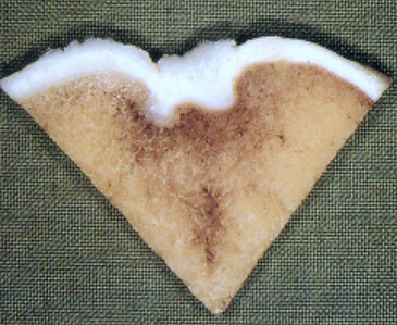


This hypothesis seems straightforward, but many concerns, including the fact that thickened cartilage or necrosis is not always seen with typical OC lesions, have been expressed that call into question the accuracy of the single hypothesis for OC. Recent investigations of the molecular mechanisms of OC indicate that the collagen component of the cartilage extracellular matrix is primarily affected. It is hypothesized that a defective collagen network results in disturbed calcification of cartilage, as has been shown in bone, causing delayed endochondral ossification, which is the hallmark of OC. Furthermore, a defective collagen network likely reduces the tensile strength of the cartilage and may, together with or without local cartilage thickening and biomechanical forces, result in the development of characteristic cartilage flaps or fragments. Osteochondrosis is a complex disease of multifactorial origin, and how these factors interrelate to produce the disease remains poorly understood.


Nutrition


Numerous investigations of the role of nutritional factors in the development of OC have been undertaken. Growth and average daily weight gain are positively correlated with OC in both the hock and stifle joints. However, it appears to be of no importance whether the high growth rate is caused by high food intake or by genetic predisposition. Overfeeding and high dietary energy intake has been shown to desynchronize several endocrine factors, including insulin, insulin-like growth factors 1 and 2 (IGF-1 and 2), and thyroid hormones tri-iodothyronine (T3) and thyroxine (T4). High dietary energy intake may desynchronize the balances among these hormones, and alterations in their secretion and metabolism may result in abnormal cartilage development.


An association between copper deficiency or zinc-induced copper deficiency and equine OC-like lesions was proposed for many years. Copper deficiency has been traditionally related to inferior collagen quality as a result of reduced cross-linking. However, more recent studies in which foals were fed less than the recommended 10 ppm of copper did not result in an increased incidence of OC. Furthermore, increased liver copper content is not related to the number of lesions, but apparently there is a positive effect of copper on the repair of lesions. High calcium concentration in the diet does not affect the occurrence of OC lesions; however, very high phosphorus concentrations increase the number of lesions. More likely, these lesions are the result of increased osteoporosis and weakening of the subchondral plate, and not OC.


Stay updated, free articles. Join our Telegram channel

May 28, 2016 | Posted by in EQUINE MEDICINE | Comments Off on Osteochondrosis

Full access? Get Clinical Tree

Get Clinical Tree app for offline access