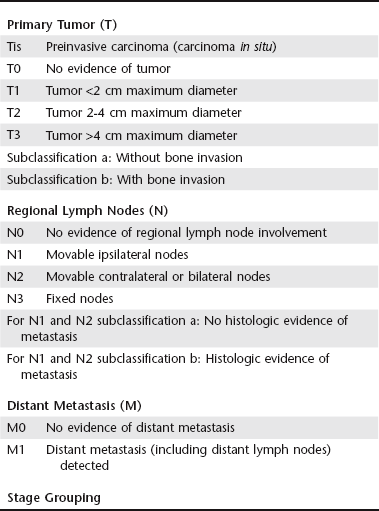
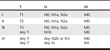
Chapter 81 Biopsy is an essential part of the evaluation of a dog or cat with an oral mass. It is not possible to distinguish tumor type or degree of malignancy based on visual inspection. Aspiration cannot often be performed without anesthesia or heavy sedation and is less likely to give a definitive diagnosis, which limits the usefulness of this test. Incisional biopsy and histopathologic evaluation provides important information regarding additional staging needed, treatment approach, and prognosis. The biopsy procedure should be planned properly so that the biopsy tract can be included easily as part of the excision of the tumor at the time of curative-intent surgery or included in the field of radiation if radiation therapy (RT) is performed. For tumors that appear locally invasive on physical examination, ideally the biopsy should be performed following imaging (see section on staging). If a definitive surgical excision is attempted, margin assessment is important to check for completeness of excision and the need for adjuvant therapy (see later). Staging of oral tumors in both dogs and cats evaluates for the extent and invasion of the local tumor as well as the presence of regional or distant metastasis. A clinical staging system has been described by the World Health Organization for use with oral tumors (Table 81-1). This staging system can be used to guide pretreatment assessment and also is important for treatment planning. Documenting the exact location and size of an oral mass helps assess the local extent of a tumor, although radiographs and computed tomography (CT) can provide more complete information as to extent and bony involvement. To assess local lymph node involvement, palpation and cytologic evaluation of mandibular lymph node aspirates is an essential first step. Because lymph node drainage is variable in the head, contrast CT studies and ultrasonography can be performed to further assess local and regional lymph nodes, which include the mandibular, parotid, retropharyngeal, and cervical lymph nodes. Ultrasonographic guidance may be necessary for sampling of all but the mandibular lymph nodes. For malignant tumors, either three-view thoracic radiography or CT of the thorax should be performed to assess for pulmonary metastasis. Benign tumors and small malignant tumors involving the mandible can be resected by mandibular rim excision. With this technique, the ventral cortex of the mandible is preserved and left intact. This avoids some of the adverse effects of a complete segmental mandibulectomy such as mandibular drift and malocclusion. Case selection for this technique is imperative. This procedure cannot be performed in a small-breed dog, and the tumor must be 10 mm dorsal to the mandibular canal. Dental radiography and CT with and without tumor contrast are necessary to ensure that a rim excision is appropriate (Arzi and Verstraete, 2010; Murray et al, 2010). If there are any doubts, it is best to perform a segmental mandibulectomy to ensure complete excision of the tumor, particularly with malignant tumors and acanthomatous ameloblastomas. Tumors of the tongue can be excised by partial or complete glossectomy in dogs. Dogs can adapt to a complete glossectomy, but a feeding tube should be placed in animals in which more than 50% of the tongue is removed because it can take several weeks for the animal to adapt (Dvorak et al, 2004). Anecdotally, cats do not do well with significant (>50%) glossectomies.
Oral Tumors
Diagnosis
Staging
Treatment—General Principles
Surgery
![]()
Stay updated, free articles. Join our Telegram channel

Full access? Get Clinical Tree


