Nematodes That Infect Domestic Animals
Learning Objectives
After studying this chapter, the reader should be able to do the following:
• Recognize pathology produced by nematodes of domesticated and laboratory animals.
• Canine roundworm (Toxocara canis)
• Canine hookworm (Ancylostoma caninum)
• Canine whipworm (Trichuris vulpis)
• Canine heartworm (Dirofilaria immitis)
• Feline heartworm (Dirofilaria immitis)
• Ruminant lungworm (Dictyocaulus viviparus)
• Equine roundworm (Parascaris equorum)
Key Terms
Prepatent period
Vermifuge
Vermicide
L2 larvae
Parthenogenesis
Microfilaria
Adulticide
Rhabditiform esophagus
Benzimidazoles
Probenzimidazoles
Strongyles
Cutaneous habronemiasis
Cutaneous draschiasis
Summer sores
Cutaneous onchocerciasis
Thumps
Asymptomatic
Oxyurids
Neotenic
This chapter includes brief descriptions of many of the common nematode parasites of domesticated (and wild) animals in the United States. The discussions include information on the tissue, organ, or organ system parasitized in the host. Where appropriate, information is also provided on the prepatent period (time from the point of infection until a specific diagnostic stage can be recovered), the diagnostic stage (life cycle stage) most often identified, a morphologic description of this diagnostic stage (e.g., eggs, larvae, or adult), and treatment for the most common parasites.
Nematodes of Dogs and Cats
Gastrointestinal Tract
Parasite: Spirocerca lupi
Host: Canines and felines
Location of Adult: Esophageal wall
Distribution: Tropical and subtropical regions
Derivation of Genus: Coiled tail
Transmission Route: Ingestion of egg
Common Name: Esophageal worm
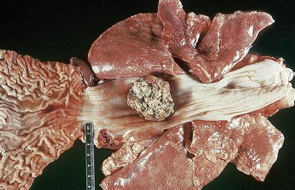
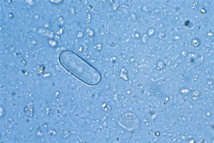
Spirocerca lupi, the esophageal worm, is a nematode usually associated with the formation of nodules in the esophageal wall of dogs and cats. Occasionally, this roundworm may be found in granulomas or nodules in the stomach wall of both dogs and cats (Figure 4-1). Adult worms reside in tunnels deep within these nodules and expel their eggs through the fistulous (tract-like) openings in the nodules. These nodules can cause obstruction of the gastrointestinal tract, esophagus, or stomach. Eggs are passed down the esophagus and out in the feces. The thick-shelled eggs of S. lupi are 30 to 37 µm ´ 11 to 15 µm and contain a larva when laid. These eggs have a unique “paper clip” shape (Figure 4-2). An animal is infected by ingesting the eggs of this parasite. Eggs usually can be observed on fecal flotation but may also be recovered from vomitus that has been subjected to standard fecal flotation procedures. Radiographic or endoscopic examination may reveal a characteristic nodule or granuloma within the esophagus or stomach. The prepatent period for this roundworm is 6 months.
 TECHNICIAN’S NOTE
Adult Spriocerca lupi reside inside nodules, which can cause esophageal obstruction.
Parasite: Physaloptera species
Host: Canines and felines
Location of Adult: Lumen of the stomach or small intestine, more commonly attached to the stomach mueosa
Distribution: Worldwide
Derivation of Genus: Bladder wing
Transmission Route: Ingestion of eggs
Common Name: Canine and feline stomach worms
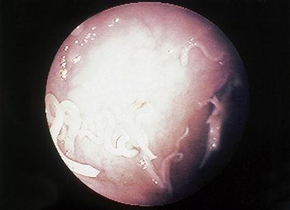
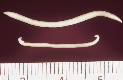
Physaloptera species are stomach worms of both the dog and the cat. Although occasionally found in the lumen of the stomach or small intestine, Physaloptera species are usually firmly attached to the mucosal surface of the stomach, where they suck blood. With the use of an endoscope, these nematodes can be observed in their attachment sites (Figure 4-3). Their diet consists of blood and tissue derived from the host’s gastric mucosa. The feeding habits of these worms may expose their attachment sites, which often continue to bleed after the parasite has detached. Vomiting, anorexia, and dark, tarry stools are often observed in infected animals. The adults are creamy white, sometimes tightly coiled, and 1.3 to 4.8 cm long (Figure 4-4). They may also be recovered in the pet’s vomitus; when this happens, Physaloptera can easily be confused with ascarids. A quick way to differentiate the two parasites is to break open an adult specimen and (if the specimen is female) examine the eggs microscopically.
The eggs of Physaloptera species are small, smooth, thick-shelled, and embryonated (larvated) when passed in feces. These eggs are quite similar in appearance to the eggs of S. lupi. They are 30 to 34 µm ´ 49 to 59 µm and contain a larva when laid. (See Figure 4-5 for the characteristic egg of Physaloptera species.) This parasite infects animals that ingest the larvated eggs. Eggs can usually be recovered on standard fecal flotation of either feces or vomitus, using solutions with a specific gravity greater than 1.25. The prepatent period for this nematode is 56 to 83 days.
 TECHNICIAN’S NOTE
The eggs of Physaloptera species can be confused with ascarids.
Parasite: Aonchotheca putorii
Host: Felines and minks
Location of Adult: Small intestine
Distribution: Tropic and temperate countries, worldwide.
Derivation of Genus: Sheath diminished in bulk
Transmission Route: Ingestion of egg
Common Name: Gastric capillarid of cats
Aonchotheca putorii is commonly referred to as the “gastric capillarid of cats.” This nematode was once known by another scientific name, Capillaria putorii. This capillarid frequently parasitizes mink but has also been reported in cats. A. putorii is rarely reported in North America. The adult parasites can be found in the small intestine where they suck blood. The eggs of A. putorii are easily confused with other trichuroid nematodes. (See the following section on identification of feline whipworms.) The eggs of A. putorii are 53 to 70 µm ´ 20 to 30 µm and exhibit a netlike surface similar to the eggs of Eucoleus aerophilus, another capillarid found in the upper respiratory system. The eggs of A. putorii are dense and less delicate than those of E. aerophilus; they have flattened sides and contain a one- or two-cell embryo that fills the egg. This parasite is passed to new hosts by ingestion of the embryonated egg. The eggs can be detected on fecal flotation.
Parasite: Ollulanus tricuspis
Host: Felines
Location of Adult: Stomach
Distribution: Worldwide
Derivation of Genus: Small feline hair
Transmission Route: Ingestion of larvae from vomitus
Common Name: Feline trichostrongyle
Ollulanus tricuspis is the feline trichostrongyle. As a group, the trichostrongyles are considered to be parasites of the gastrointestinal tract of ruminants, such as cattle, sheep, and goats; it is quite unusual for the cat to serve as a host for a trichostrongyle. O. tricuspis is often diagnosed in cats that exhibit chronic vomiting. These nematodes are usually identified by examining the cat’s vomitus under a dissecting or compound microscope. Feline vomitus can also be examined with a standard fecal flotation procedure. The best flotation solution for identification is a modified Sheather’s flotation solution. Adult female O. tricuspis are tiny, only 0.8 to 1 mm long, and possess three major tail cusps, or toothlike processes, on the tip of the tail (thus the specific epithet tricuspis). Adult male nematodes are 0.7 to 0.8 mm long and possess a copulatory bursa. The female worms are larviparous and release infective third-stage larvae (500 × 22 µm), which mature to adults in the cat’s stomach. This feline trichostrongyle also is unusual in that these third-stage larvae are immediately infective for any cat that may ingest vomitus containing them. Free-living stages in the external environment are not required for completion of the life cycle.
 TECHNICIAN’S NOTE
Ollulanus tricuspis females release third-stage infective larvae that are immediately infective to cats.
Parasites: Toxocara canis, Toxocara cati, and Toxascaris leonina
Host: Canines and felines
Location of Adults: Small intestine
Distribution: Worldwide
Derivation of Genus: Toxocara – arrowhead, Toxascaris – arrow ascaris
Transmission Route: Ingestion of egg with infective larva
Common Name: Canine and feline roundworms
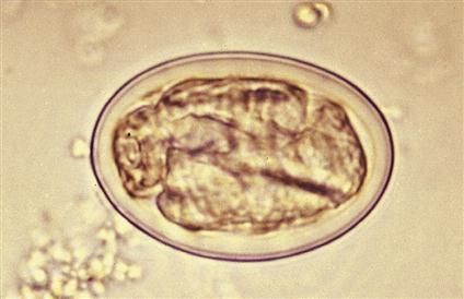
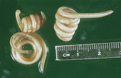
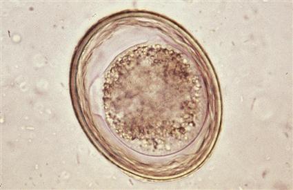
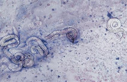
Toxocara canis, Toxocara cati, and Toxascaris leonina are the ascarids, or large roundworms, of dogs and cats. These roundworms may be found in the small intestine of dogs and cats in most areas of the world. All young puppies and kittens presenting to a veterinary clinic should be examined for the presence of these large, robust nematodes. Adult ascarids may vary in length from 3 to 18 cm and, when passed in the feces, are usually tightly coiled, similar to a coiled bedspring (Figure 4-6). Ascarids in young puppies and kittens may produce vomiting, diarrhea, constipation, and other nonspecific clinical signs. The adult worm does not attach to the host but rather uses an undulating motion to remain in the small intestine. Therefore, adult worms may “swim” into the stomach and cause vomiting. The worm may then be present in the vomitus. Because of the presence of the adult worm in the vomitus, the owner may describe the vomitus as having “spaghetti” in it. An accumulation of adults can cause gastrointestinal obstruction.
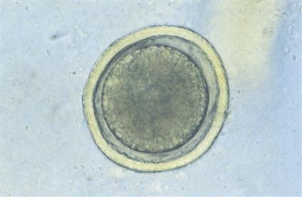
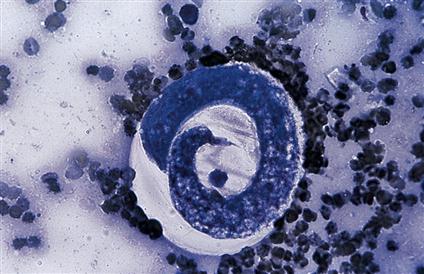
The eggs of Toxocara species are unembryonated, spherical, and have a deeply pigmented center and rough, pitted outer shell. Eggs of Toxocara canis are 75 ´ 90 µm in diameter (Figure 4-7), whereas those of Toxocara cati are smaller, 65 to 75 µm in diameter (Figure 4-8). The eggs of Toxascaris leonina are spherical to ovoid, with dimensions of 75 ´ 85 µm. In contrast to the eggs of Toxocara species, the eggs of T. leonina have a smooth outer shell and a hyaline, or “ground glass,” central portion (Figure 4-9). The prepatent period for T. canis is 21 to 35 days, whereas that of T. leonina is 74 days. Ascarids are among the most frequently diagnosed nematodes in young puppies and kittens.
 TECHNICIAN’S NOTE
Adult ascarids are commonly seen in vomitus and may be described by pet owners as “spaghetti-like” worms.
Adult roundworms are present in the small intestine, where they mate. The female produces unembryonated eggs that are passed in the host’s feces. The eggs embryonate on the ground, to the point that they contain L2 larvae. They are ingested by a host and the L2 larvae are released from the eggs. The L2 larvae may go into dormancy in an adult host, but they grow and migrate to various tissues in the young host. Finally, the L2 larvae migrate to the young host’s lungs, where they are coughed up and swallowed by the host. They grow to adulthood in the host’s small intestine and begin a new life cycle (Figure 4-10).
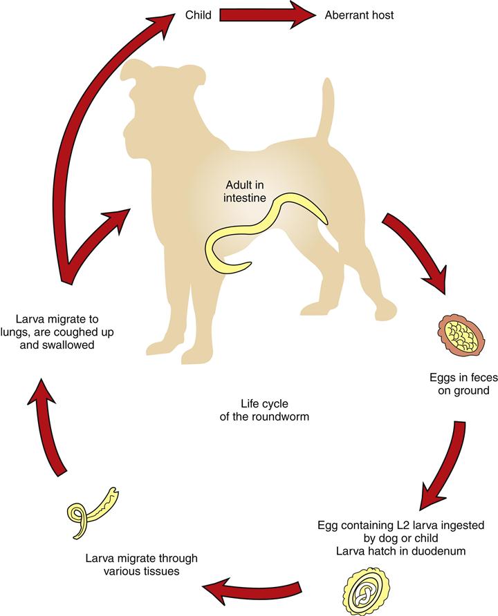
In the adult female host, the infective larvae remain dormant until the female host mates and produces hormones during pregnancy. At this time the larvae become active once more and migrate through the female host’s body. The larvae (with the exception of T. cati) can cross the placental barrier to infect the host’s offspring. The most common symptoms of a roundworm infection are diarrhea, vomiting, and a pot-bellied appearance.
The most common way of identifying an infection by T. canis, T. cati, or T. leonina is through the fecal flotation technique. Usually the pregnant female will have feces tested for these parasites to determine the chances of her puppies or kittens becoming infected. Puppies and kittens are routinely dewormed during their first few visits to the veterinary office.
Several types of anthelmintics can be used to treat a roundworm infection. A vermifuge (anthelmintic that paralyzes the parasite so it passes out in the feces) such as piperazine or pyrantel is often used to treat this type of parasite. A vermicide (anthelmintic that kills the parasite and allows the parasite to be broken down by the body) such as thiabendazole or mebendazole can also be used to treat a roundworm infection.
 TECHNICIAN’S NOTE
The best way to identify a roundworm infection is through the fecal flotation technique.
T. canis, although primarily a parasite of the dog, can also infect humans (visceral larva migrans), as discussed in Chapter 17. The best way to prevent infection to humans and other animals is cleaning up feces in the yard daily. Also, prevent children from playing in the yard and placing their contaminated hands to their mouths.
Parasite: Baylisascaris procyonis
Host: Raccoon, but also seen in many species
Location of Adult: Small intestine
Distribution: North America (Mississippi-Ohio River basin)
Derivation of Genus: Named after a famous parasitologist, Dr. Baylis
Transmission Route: Ingestion of eggs
Common Name: Raccoon roundworm
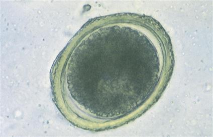
Baylisascaris procyonis is the roundworm of the raccoon. Baylisascaris procyonis is common in North America and is a zoonotic parasite. The adult resides in the small intestine of the raccoon. The ova are found in the feces. The ova are unembryonated and spherical with a deeply pigmented center and a rough/pitted outer shell. It is slightly smaller than the ova of Toxocara canis (Figure 4-11). Accumulation of the adults in the small intestine can cause a gastrointestinal obstruction in the natural host.
B. procyonis is diagnosed by finding the ova on fecal flotation. The ova can be found anywhere that raccoons defecate and in naturally infected canines. While the raccoon is the natural host, it can cause zoonotic disease in many species. Since the larvae migrate to the central nervous system, this is a very pathogenic parasite in the incidental host (Bowman, 2009), producing a zoonotic condition known as neurologic larva migrans
 TECHNICIAN’S NOTE
Baylisascaris procyonis is a very pathogenic zoonotic parasite that can infect humans.
Parasites: Ancylostoma caninum, Ancylostoma tubaeforme, Ancylostoma braziliense, and Uncinaria stenocephala
Host: A. caninum (canine), A. tubaeforme (feline), A. braziliense (canine and feline), and U. stenocephala (canine)
Location of Adult: Small intestine
Distribution: Ancylostoma species (worldwide), Uncinaria stenocephala (northern regions of North America)
Derivation of Genus: Ancylostoma-curved mouth and straight trumpet shape, Uncinaria-hooked nose and narrow head
Transmission Route: Ingestion of eggs, through the skin, across placenta, and through mammary milk
Common Name: Canine and feline hookworm
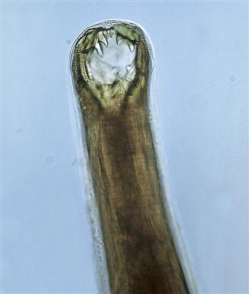
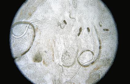
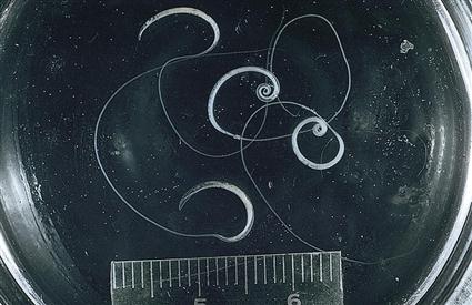
Ancylostoma caninum, the canine hookworm; Ancylostoma tubaeforme, the feline hookworm; Ancylostoma braziliense, the canine and feline hookworm; and Uncinaria stenocephala, the northern canine hookworm, are nematodes that infect the small intestine. Hookworms are found throughout the world but are most often found in tropical and subtropical areas of North America. Hookworms attach to the small intestinal mucosa. A. caninum demonstrates a frightful buccal cavity—it has three pairs of ventral teeth with which it attaches to the mucosa of the small intestine (Figure 4-12). These parasites may change feeding sites and reattach elsewhere in the small intestine. Because this hookworm feeds on blood, it secretes an anticoagulant from its mouth, which causes the former attachment sites to continue to bleed. Thus the hookworm’s voracious feeding activity and the secondary hemorrhage produced can cause significant anemia. Because this bleeding occurs within the small intestine, the blood is often digested by the host and often appears as a black, tarry stool. The resulting anemia can be quite severe in young kittens and puppies. Hookworms often produce serious problems in kennels and catteries. The prepatent period depends on the species of hookworm. Animals are usually infected through percutaneous, prenatal, and transmammary routes.
The male and female worms attach to adjacent villi of the intestinal wall. The male and female mate continuously, and therefore the female continually produces eggs that are laid in the feces. The egg contains the morula stage as it is passed out of the host’s body in the feces. These eggs embryonate into L1 larvae in the external environment. The L1 larvae hatch, feed, grow, and molt to L2 larvae. The L2 larvae feed, grow, and molt to L3 larvae, but do not release the cuticle of the L2 larvae. They are ensheathed L3 larvae that do not feed. They are the infective stage larvae. The L3 larvae can be ingested, or they can penetrate the host’s intact skin and migrate through the tissues of the body until they reach the lungs. Once in the lungs, the larvae are coughed up and swallowed by the host (Figure 4-13).
 TECHNICIAN’S NOTE
Hookworms can cause a black, tarry stool in the host due to the attachment and reattachment of the parasite and digestion of free blood in the intestinal tract.
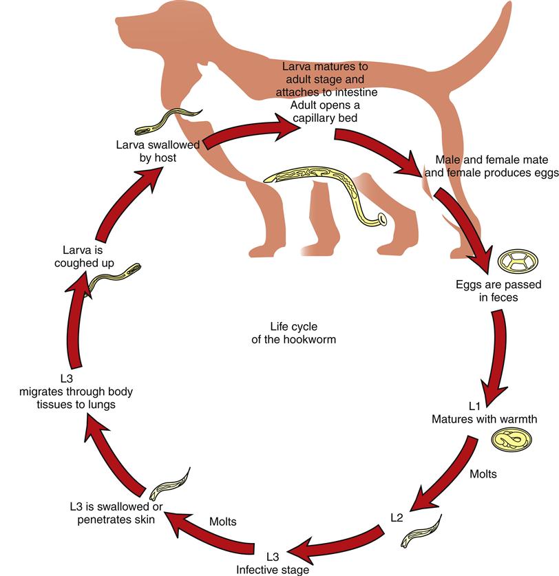
The larvae of this parasite are able to cross the placenta and also can be passed from mother to offspring through the colostrum. Therefore the pregnant host should be checked several times during pregnancy for the presence of these parasites. Since these parasites can penetrate intact skin, they can also enter the skin of humans (cutaneous larval migrans), as discussed in Chapter 17.
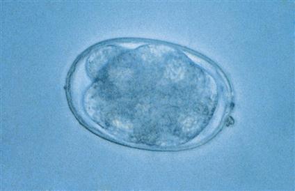
Eggs of all hookworm species are oval or ellipsoidal, thin-walled, and contain an 8- to 16-cell morula when passed in feces. Because these eggs embryonate, or larvate, rapidly in the external environment (as early as 48 hours), fresh feces are needed for diagnosing hookworm infections. Eggs of A. caninum are 56 to 75 µm × 34 to 47 µm; those of A. tubaeforme, 55 to 75 µm × 34.4 to 44.7 µm; those of A. braziliense, 75 × 45 µm; and those of U. stenocephala, 65 to 80 µm × 40 to 50 µm. These eggs can be easily recovered on standard fecal flotation (Figure 4-14).
 TECHNICIAN’S NOTE
Hookworms are easily recovered and identified in a standard fecal flotation technique.
Treatment for the hookworm species can take two forms. The first is prevention with the use of once-a-month heartworm preventive agents; Interceptor™ and Heartgard Plus™ are the most common brands although there are other brands of heartworm preventatives that also work against hookworms. Many of these preventive medications are also labeled for the prevention of hookworms. If the parasite eggs are found on fecal flotation or other techniques, the veterinarian may choose a vermicide such as mebendazole or fenbendazole.
The best way to prevent hookworm species from infecting animals or humans is removing the feces after defecation by the host. Removal during warm weather should take place immediately after defecation, whereas removal during cold months may take place every other day.
 TECHNICIAN’S NOTE
The best way to prevent hookworm infections is to remove feces from the yard on a daily basis before the larvae hatch from the ova.
Parasites: Strongyloides stercoralis and Strongyloides tumiefaciens
Host: Dogs, cats, and humans
Location of Adult: Small intestines
Distribution: Worldwide
Derivation of Genus: Round-like shape
Transmission Route: Through the skin and in mammary milk
Common Name: Intestinal threadworms
Because parasitic males do not exist, the female worms are capable of producing viable ova without fertilization from a male worm. This process is called parthenogenesis.
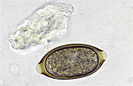
Strongyloides stercoralis and S. tumiefaciens are often referred to as intestinal threadworms. These nematodes are unique in that only a parthenogenetic female is parasitic in the host and resides in the small intestines. The female has an approximately cylindric esophagus that is one-quarter the length of the parasite’s body. These females produce embryonated or larvated eggs, but in dogs, the eggs hatch in the intestine, releasing first-stage larvae that may be observed in fresh feces. (Figure 4-15 shows the parasitic adult females, eggs, and first-stage larvae of Strongyloides species.) The larval Strongyloides species are 280 to 310 mm long and possess a rhabditiform esophagus with a club-shaped anterior corpus, a narrow median isthmus, and caudal bulb. The larvae go through a free-living stage in the environment before becoming infective third-stage larvae that can penetrate the skin and cause migratory damage in the host. In the dog, the major method of transmission is from mother to pups through the mammary glands. The prepatent period is 8 to 14 days. These nematodes are frequently associated with a moderate-to-severe diarrhea in young puppies, particularly in kennel environments during the summer months. Since humans can also be infected with S. stercoralis there is the potential of zoonosis called Strongyloidiasis.
 TECHNICIAN’S NOTE
In dog feces, you will commonly find the larvae (rather than ova) of Strongyloides species.
Parasites: Trichuris vulpis, Trichuris campanula, and Trichuris serrata
Host: T. vulpis (canine), T. campanula, and T. serrata (feline)
Location of Adult: Cecum and colon
Distribution: Worldwide but T. campanula and T. serrata are rare in North America
Derivation of Genus: Hair tail
Transmission Route: Ingestion of eggs
Common Name: Whipworm
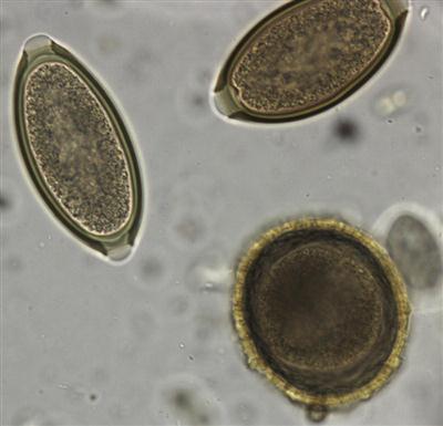
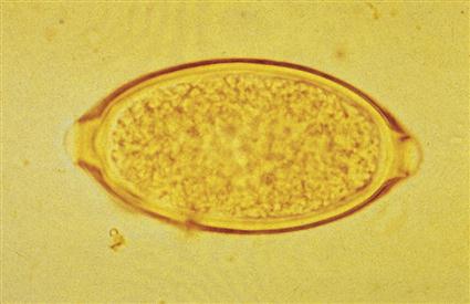
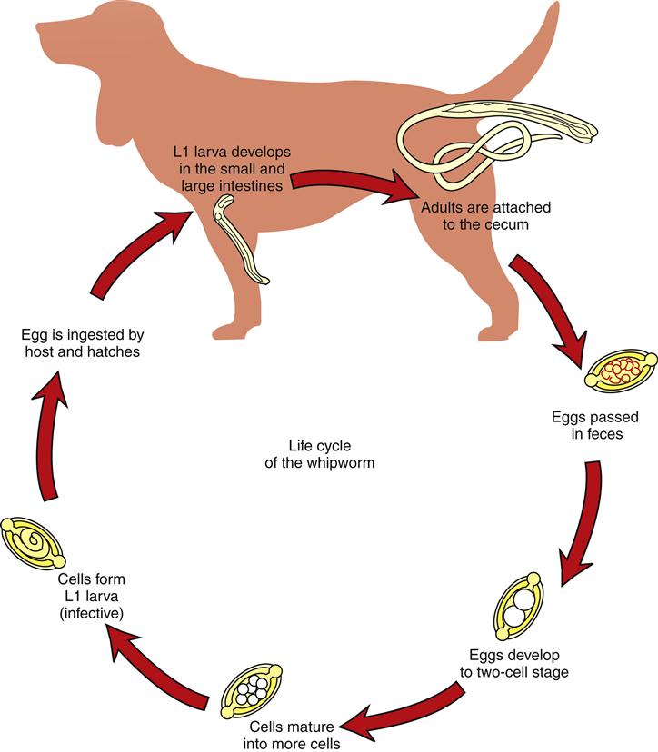
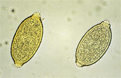
Trichuris vulpis, the canine whipworm, and Trichuris campanula and Trichuris serrata, the feline whipworms, reside in the cecum and colon of their respective definitive hosts. Whipworms are a common clinical occurrence in dogs; however, it is important to remember that feline whipworms are quite rare in North America and have been diagnosed only sporadically throughout the world. Whipworms derive their name from the fact that the adults have a thin, filamentous anterior end (the “lash” of the whip) and a thick posterior end (the “handle” of the whip (Figure 4-16). The thin end of the whipworm was once thought to be the tail of the parasite. The egg of the whipworm is described as trichuroid or trichinelloid; it has a thick, yellow-brown, symmetric shell with prominent polar plugs at both ends. The eggs are unembryonated (not larvated) when laid. Eggs of T. vulpis are 70 to 89 mm ´ 37 to 40 mm. (Figure 4-17 shows the characteristic egg of T. vulpis.) The prepatent period for T. vulpis is 70 to 90 days.
The adult Trichuris species are attached to the wall of the cecum or colon where they suck blood and produce eggs. The eggs are passed out in the feces of the host every third day. Once in the environment, the eggs will go through a period of development to become an L2 (infective) larva within the egg. Unlike the hookworm species, the whipworm species must be ingested by the host. The L2 larva will hatch from the egg once in the host’s small intestine. The larva will molt several times in the small and large intestine before becoming an adult. The new adult will migrate back through the colon to the cecum and attach to the intestinal wall (Figure 4-18). The most common symptoms seen in infected animals are diarrhea, anemia, and mucus-coated stool.
 TECHNICIAN’S NOTE
The eggs of Trichuris species are only passed in the stool every third day rather than every day like most nematodes.
The eggs of T. campanula and T. serrata, the feline whipworms, may be easily confused with Aonchotheca putorii, Eucoleus aerophilus, and Personema feliscati, parasites of the feline stomach, respiratory tract, and urinary system, respectively. The eggs of T. campanula average 63 to 85 µm ´ 34 to 39 µm (Figure 4-19). When examining a cat’s feces for trichurids, it is important that the diagnostician remember that pseudoparasites, that is, the eggs of trichurids or capillarids, may parasitize nonfeline hosts, such as mice, rabbits, or birds (an outdoor cat’s prey). The eggs of trichurids or capillarids from a cat’s prey may pass undigested and unaltered through the cat’s gastrointestinal system, remaining intact and unembryonated.
Fecal flotation is the most common way of identifying the eggs of this parasite; however, whipworm eggs do not float well in most common fecal flotation media. Therefore, it is imperative that the eggs be allowed to float for a minimum of 15 minutes before viewing under the microscope. Once identified, the veterinarian will most often use the vermicides mebendazole or fenbendazole for treatment of these worms. Prevention involves daily feces removal to remove the uninfective eggs from the host’s environment.
Parasite: Enterobius vermicularis
Host: Humans
Distribution: Worldwide
Derivation of Genus: Intestinal life
Transmission Route: Ingestion of eggs
Common Name: Human pinworm
Dogs and cats may often be blamed for serving as hosts for certain parasites of humans. Enterobius vermicularis is the pinworm found in humans and is often found in young children. This pinworm does not parasitize dogs or cats. Nevertheless, the family pet is often falsely incriminated by physicians, family practitioners, and pediatricians as a source of pinworm infection in young children. The veterinary diagnostician should remember this rule: Pinworms are parasites of omnivores (mice, rats, monkeys, and humans) and herbivores (rabbits and horses) but never carnivores (dogs and cats).
 TECHNICIAN’S NOTE
Because whipworm ova are heavy and hard to float, they require at least 15 minutes with a standard fecal flotation technique before viewing under a microscope. Ideally, centrifugal flotation should be used to better float the whipworm ova.
Circulatory System
Parasite: Dirofilaria immitis
Host: Canine, feline, and ferret. Has been observed in humans as an incidental parasite.
Intermediate Host: Female mosquito
Location of Adult: Right ventricle and pulmonary arteries
Distribution: Warm-temperate climates around the world
Derivation of Name: Dread thread and inexorable
Transmission Route: Bite of infective mosquito
Common Name: Heartworm
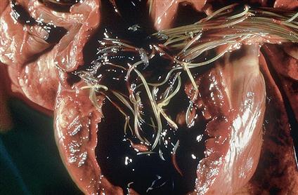
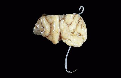
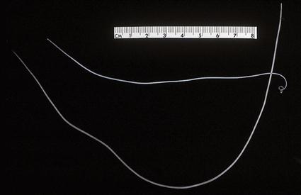
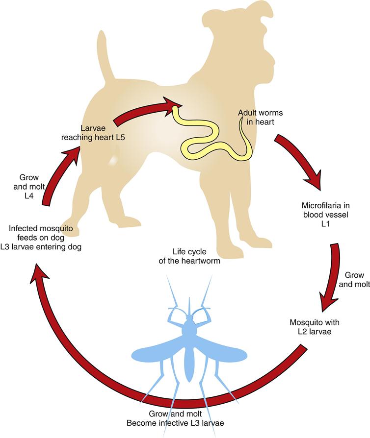
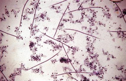
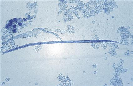
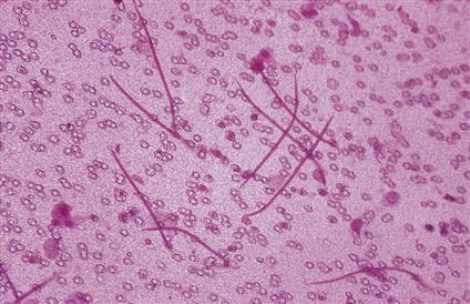
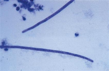
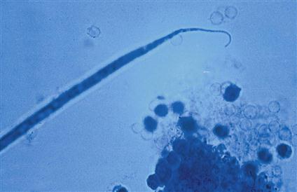
Dirofilaria immitis is often referred to as the canine heartworm (Figure 4-20); however, this nematode has been known to parasitize cats and ferrets as well (Figure 4-21). The definitive host for D. immitis is the canine. Although cats and ferrets have been parasitized by this parasite, they rarely serve as a source for transmission. Heartworms are long, slender parasites (Figure 4-22). As adults, these parasites are found within the right ventricle and the pulmonary artery and its fine branches. The offspring of filarial worms such as D. immitis are known as microfilariae.
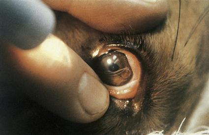
Among the nematodes of small animals, D. immitis is perhaps the one nematode that may be found in sites other than its normal predilection sites. This parasite is often recovered in a variety of aberrant sites, such as the brain, anterior chamber of the eye (Figure 4-23), and subcutaneous sites. The prepatent period in dogs is approximately 6 months.
 TECHNICIAN’S NOTE
Dirofilaria immitis is the parasite that causes heartworm disease in dogs and cats. It has a prepatent period of 6 months before it can be diagnosed in the dog.
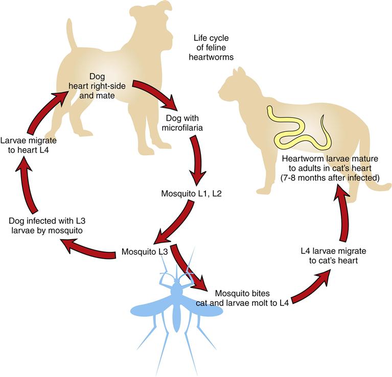
The life cycle of D. immitis requires the mosquito intermediate host to be transmitted from animal to animal. The adults live in the right ventricle and pulmonary artery where they can obstruct the blood vasculature. The male and female adults mate, and the female produces microfilariae. The microfilariae are released into the host’s bloodstream, where they are ingested by feeding female mosquitoes. The microfilariae grow and molt in the mosquito until they reach the infective stage. Once they become infective, they enter a new host the next time the mosquito feeds (Figure 4-24). Once in the new host, the larvae migrate and molt through various body tissues on their way to the heart. It is at this time that the larvae may grow and molt to become adults in sites other than the heart.
 TECHNICIAN’S NOTE
Dirofilaria immitis larvae must develop to an infective stage within a mosquito before they can be transferred to a new host.
In microfilaremic dogs, diagnosis is made by observing microfilariae in blood samples, using one of several concentration techniques, such as the modified Knott’s test (Figures 4-25 and 4-26) or commercially available filter techniques (Figure 4-27). For dogs (amicrofilaremic or microfilaremic), infection can also be diagnosed using one of the commercially available enzyme-linked immunosorbent assay (ELISA) tests (Figure 4-28). It is important to remember that a subcutaneous filariid of dogs, Acanthocheilonema (formerly known as Dipetalonema) reconditum, also produces microfilariae in the peripheral blood (Figures 4-29 and 4-30). The microfilariae of this nonpathogenic nematode must be differentiated from those of D. immitis (see Figures 4-25 and 4-26).
The damage that D. immitis causes to the heart will cause the hallmark symptoms of heartworm disease in dogs. The symptoms are a decrease in exercise tolerance, right-sided heart enlargement, and abdominal ascites (fluid accumulation in the abdomen). These symptoms are caused by the parasite’s location in the heart and pulmonary arteries, which reduces blood flow on the right side of the heart and causes inflammation to the lining of the blood vessels.
 TECHNICIAN’S NOTE
The classic symptoms of heartworm disease are exercise intolerance, right-sided heart enlargement, and abdominal ascites caused by reduced blood flow on the right side of the heart.
The treatment for heartworm disease involves pretreatment testing, treatment, and posttreatment rest. The first step in treatment involves determining the canine’s ability to withstand the treatment by performing blood work; this determines the status of the internal organs. Radiographs are taken to determine the status of the heart and the stage of the disease. After testing, the canine is treated for the adult heartworms. Adulticides (drugs that will kill the adult stages of the parasite) such as Immiticide™ (Merial Limited, www.merial.com) are used to kill the adult heartworms. As the adults die, they will move with the flow of blood toward the lungs. This could cause problems for the canine if it is allowed to exercise after the adulticide treatment. The canine must be kept quiet for several weeks after treatment while the body resorbs the dead adults. Once the adults have been treated and resorbed by the body, the canine is treated with a microfilaricide (usually ivermectin) to clear the blood of any microfilariae. The final part of the treatment is the heartworm testing. This usually involves a microfilariae test and an ELISA test to confirm the microfilariae and adults have been cleared from the canine’s body.
The treatment for canine heartworm disease can be very stressful on the dog’s body. Therefore, prevention is recommended so that the dog does not contract the parasite. The various preventive regimens include daily preventive medications, monthly medications, and an injection every 6 months.
Feline hosts for Dirofilaria immitis occur in the same areas that canines are infected. Cats are susceptible to the parasite but tend to be very resistant to infection. Therefore, it takes a greater exposure to the microfilariae from the mosquito to infect a cat than it does a dog. Whereas D. immitis causes enough damage to produce hallmark symptoms in dogs, cats do not show specific symptoms of the infection. Cats tend to show signs similar to those seen with respiratory disease.
The life cycle of D. immitis in cats begins as it does in dogs. The adults live in the right ventricle and pulmonary arteries of the dog. The adults mate, and the female produces microfilariae, which are present in the vessels of the dog. Mosquitoes feed on the infected canine and ingest the microfilariae. The microfilariae grow and molt to the infective L3 larvae. Once infective, the mosquito feeds on a cat, and the larvae are transferred to the cat. The larvae grow, molt, and migrate in the cat on their way to the heart (Figure 4-31).
 TECHNICIAN’S NOTE
Feline heartworm diseases do not produce the hallmark symptoms observed in canine heartworm disease. The symptoms are similar to those seen with respiratory disease.
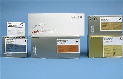
Identification of the parasite in the cat is difficult. The adults do not produce many microfilariae in the cat, so the microfilarial tests will produce negative results in most cases. Tests developed for dogs are not sensitive enough for the infection in cats, so ELISA tests have been developed for detection of D. immitis in cats. Two types of tests are available for in-clinic use: antigen tests and antibody tests. The antigen test is the definitive test, although a negative result does not rule out a heartworm infection. The antibody test can detect a reaction by the cat’s body to the microfilariae, but the microfilariae may die before becoming adults. Therefore the positive antibody test does not positively detect an active infection. A positive antigen or antibody test in the presence of clinical respiratory symptoms indicates an active infection (www.heartwormsociety.org/felineheartworminfo).
At this time there are no approved treatments for feline heartworm disease. Therefore the best treatment is prevention. Preventive options available for cats include once-a-month pills or a single topical monthly application for the prevention of feline heartworm disease.
 TECHNICIAN’S NOTE
The best way to test for feline heartworm disease is to use both an antigen test and an antibody test made for feline heartworm disease since those made for canines alone are not sensitive enough for feline heartworm disease.
Respiratory System
Parasite: Aelurostrongylus abstrusus
Host: Feline
Location of Adult: Respiratory bronchioles and alveolar ducts
Distribution: Worldwide
Derivation of Genus: Cat strongyle
Transmission Route: Ingestion of larvae
Common Name: Feline lungworm
Aelurostrongylus abstrusus is the feline lungworm. The adults live in the terminal respiratory bronchioles and alveolar ducts, where they form small egg nests or nodules where they can obstruct the bronchioles. Initially, this parasite lays embryonated (larvated) eggs. The eggs of this parasite are forced into the lung tissue, where they hatch to form characteristic first-stage larvae, approximately 360 mm long. These larvae are quite distinct; each has a tail with a unique, S-shaped bend with a dorsal spine (Figure 4-32). These larvae are coughed up by the cat, swallowed, and passed out in the feces. Diagnosis is made by finding these characteristic larvae on fecal flotation or by the Baermann technique (see Chapter 17). It is also possible to recover the larvae using a tracheal wash (Figure 4-33). The prepatent period for the feline lungworm is approximately 30 days.
Parasite: Filaroides osleri, Filaroides hirthi, and Filaroides milksi
Host: Canine
Location of Adult: Trachea, lung parenchyma, and bronchioles, respectively
Distribution: North America, Europe, and Japan
Derivation of Genus: Thread-like shape
Transmission Route: Ingestion of infected L1 larvae
Common Name: Canine lungworm
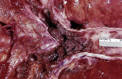
Filaroides osleri, F. hirthi, and F. milksi, the canine lungworms, are found in the trachea, lung parenchyma, and bronchioles, respectively. The larva is 232 to 266 µm in length and has a tail with a short, S-shaped appendage. Filaroides species are unique among nematodes in that their first-stage larvae are immediately infective for the canine definitive host. No period of development is required outside the host. These parasites are transmitted from the dam to her offspring as she licks and cleans her puppies. Diagnosis is by finding these characteristic larvae on fecal flotation or by the Baermann technique. (Figure 4-34 shows the unique infective larvae of F. osleri.) Nodules of F. osleri are usually found at the bifurcation of the trachea, where they can cause obstruction of the airway and be easily observed at necropsy (Figure 4-35). The prepatent period for F. osleri is approximately 10 weeks.
Parasite: Eucoleus aerophilus and Eucoleus böehmi
Host: Canines/felines and canines, respectively
Location of Adult: Bronchi and trachea, nasal cavity and frontal sinuses, respectively
Distribution: North America, South America + Europe
Derivation of Genus: Good sheath
Transmission Route: Ingestion of ova from feces or mucoid discharge
Common Name: Respiratory capillarid
Eucoleus aerophilus (Capillaria aerophila) is a capillarid nematode found in the trachea and bronchi of both dogs and cats where they can cause an inflammatory response by the host. The prepatent period is approximately 40 days. The ova are passed through the feces or mucoid discharge from coughing them up in sputum. The ova are ingested by the host. During standard fecal flotation, eggs of Eucoleus species are often confused with those of Trichuris species (whipworms). Eggs of E. aerophilus are smaller than whipworm eggs (59 to 80 µm × 30 to 40 µm), more broadly barrel-shaped, and lighter in color. The egg has a rough outer surface with a netted appearance (Figure 4-36). Eucoleus böehmi is found in the nasal cavity and frontal sinuses of dogs where it can cause obstruction of the nasal cavity and sinuses. Its eggs are smaller and have a smoother outer surface than those of E. aerophilus. Its shell has a pitted appearance. E. böehmi can be diagnosed by standard fecal flotation.
 TECHNICIAN’S NOTE
Eucoleus aerophilus and Eucoleus böehmi ova are easily confused with Trichuris ova on fecal flotation.
Urinary Tract
Parasite: Dioctophyma renale
Host: Canine
Location of Adult: Right kidney
Distribution: North America (primarily Canada) and Europe (but not Britain)
Derivation of Genus: Distended growth
Transmission Route: Ingestion of infected larvae within annelid
Common Name: Canine giant kidney worm
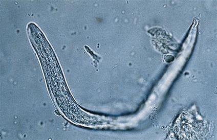
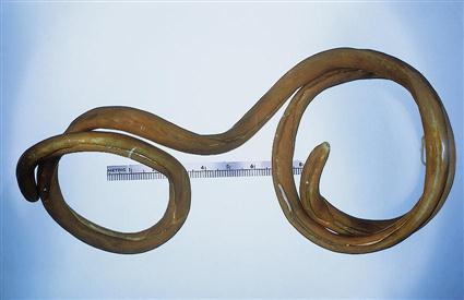
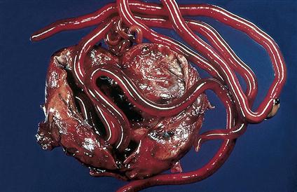
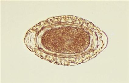
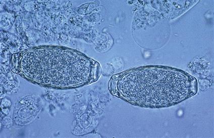
Dioctophyma renale is the giant kidney worm of dogs. This largest of the parasitic nematodes that affect domestic animals (Figure 4-37) frequently infects the right kidney of dogs and ingests the parenchyma, leaving only the capsule of the kidney (Figure 4-38). The eggs from the canine urine will be ingested by an annelid worm and mature into an infective larva. Ingestion of the annelid worm with an infective larva will spread the parasite to a new canine host. Eggs may be recovered by centrifugation and examination of the urine sediment. They are characteristically barrel-shaped, bipolar, and yellow-brown. The shell has a pitted appearance. Eggs measure 71 to 84 µm × 46 to 52 µm. (Figure 4-39 shows the characteristic egg of D. renale.) D. renale is also known for its wandering activity; it often “gets off course” during its migration to the kidney. This nematode also may occur freely within the peritoneal cavity. When the kidney worm is in an aberrant site, its eggs cannot be passed to the external environment. The prepatent period for this largest of nematodes is approximately 18 weeks.
 TECHNICIAN’S NOTE
Dioctophyma renale is the largest parasitic nematode of domestic animals and can ingest the parenchyma of the right kidney.
Parasite: Pearsonema plica and Pearsonema feliscati
Host: Canines and felines, respectively
Location of Adult: Urinary bladder
Distribution: Southeastern US
Transmission Route: Ingestion of infective intermediate host, earthworm; ingestion of earthworm containing infected L3 larvae
Common Name: Bladder worm of canines and felines, respectively
Pearsonema plica and Pearsonema feliscati (formerly capillaria) are nematodes of the urinary bladder of dogs and cats, respectively. Their eggs may be found in urine or in feces contaminated with urine. Eggs are clear to yellow, measure 63 to 68 µm × 24 to 27 µm, and have flattened, bipolar end plugs. The outer surface of the shell is roughened (Figure 4-40). These eggs may be confused with eggs of the respiratory and gastric capillarids and eggs of the whipworms. The eggs are ingested by an earthworm where they mature to infective larvae. If the earthworm is ingested by a new host, the larvae will migrate to the urinary bladder.
 TECHNICIAN’S NOTE
Pearsonema species can be found in urine sediment or in fecal samples contaminated with urine but the eggs can be easily confused with other capillarid ova of the respiratory and intestinal tract as well as with Trichuris species ova.
Stay updated, free articles. Join our Telegram channel

Full access? Get Clinical Tree


