Leptospirosis

CHAPTER 33 Leptospirosis


Leptospirosis is caused by highly invasive, spiral bacteria of the genus Leptospira. The infectious agent is capable of infecting both humans and animals. Less is known about leptospirosis in horses than in any of the common domestic animals except cats. On the basis of DNA-DNA reassociation studies, genus Leptospira is classified into 13 named species and 4 genomospecies, several of which contain both pathogenic and nonpathogenic serovars. Serovars, which are based on the older phenotype classification, are sometimes classified as causing host-adapted infection or incidental host infection. Host-adapted strains seldom cause clinical disease in their maintenance host, infection and shedding are prolonged, and the serologic response following infection is relatively low. Conversely, incidental host serovars are more likely to cause clinical disease in a nonmaintenance host, a marked serologic response occurs following infection, and there is only a brief period of shedding.


In North American horses, Leptospira interrogans serovar Pomona type kennewicki is the prominent incidental (pathologic) serovar, and the skunk is the most common maintenance host of this serovar. In Europe, important equine strains are Leptospira kirschneri serovar Grippotyphosa, strains duster (western Europe) and moskva (eastern Europe). L. interrogans serovar Bratislava is considered by most researchers to be the host-adapted serovar of the horse. This belief is met with some controversy, however, as horses may have high serum titers of antibodies against serovar Bratislava, and some investigators believe it to be pathogenic in horses.


CLINICAL SYNDROMES


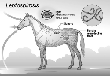
Pathogenic Leptospira infections in the horse appear to have organ trophism for the kidney, eye, or female reproductive tract (Figure 33-1). Infection may result in placentitis and abortion, acute renal failure or hematuria, and, importantly, uveitis.


Reproductive Tract Infection


L. interrogans serovar Pomona abortions account for approximately 13% of bacterial abortions in mares in endemic regions. Serovar Pomona is responsible for most of the Leptospira abortions in North America, but serovars Grippotyphosa and Hardjo have also been reported. Most abortions occur after 9 months of gestation, and, rarely, a live foal may be born ill from leptospirosis. Moreover, infected fetuses carry Leptospira in the placenta, umbilical cord, kidney, and liver. Lesions include placentitis that does not involve the cervical star. Macroscopic lesions are edema and areas of necrosis in the chorion. Microscopic lesions include necrosis and calcification of the placenta. Macroscopically the fetal liver may have yellow discoloration. Liver disease is caused by multifocal necrosis and giant cell hepatopathy. Tubulonephrosis and interstitial nephritis may be present in the kidney of the aborted fetus. Inflammation of the umbilical cord, funisitis, may be recognized by diffuse yellowish discoloration. It is unknown whether abortion results because of the placentitis, funisitis, or fetal infection or the effects of all three. Although more than one mare on a farm may abort because of Leptospira infection, abortions in epidemic areas are rare. Aborting mares and other recently infected horses are believed to shed L. interrogans serovar Pomona in the urine for approximately 2 to 3 months. A small number of horses on a farm with one or more Leptospira abortions may develop uveitis weeks later.


Stay updated, free articles. Join our Telegram channel

May 28, 2016 | Posted by in EQUINE MEDICINE | Comments Off on Leptospirosis

Full access? Get Clinical Tree

Get Clinical Tree app for offline access