6 Large eyelid mass
CLINICAL EXAMINATION
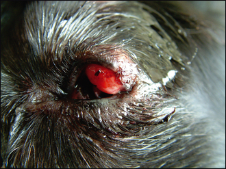
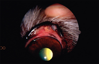
General clinical examination is normally unremarkable, although occasionally a localized lymphadenopathy is present and should be checked for. On ophthalmic examination a large mass can be seen on either the upper or lower lid margin. The periorbital skin might be thickened and inflamed. The mass itself will probably involve the lid margin, but not invariably so (Figure 6.1). The mass might be smooth or lobulated and can vary in colour from pale pink through red to darkly pigmented. The palpebral conjunctiva will be inflamed and the lid margin should always be everted to check the internal extent of the mass – frequently what can be seen from the outside is just the ‘tip of the iceberg’ (Figure 6.2). Some ocular discharge is normally present – this might be serous but is commonly mucopurulent. In some patients it will be excessive and can stick the eyelids together. Schirmer tear test is usually normal. The cornea should be carefully examined – some keratitis is common where the lid mass rubs on the cornea during blinking, and ulceration is not uncommon. Intraocular examination is usually unremarkable, although there might be slight miosis, representing a reflex uveitis if significant trigeminal pain is present.
Table 6.1 Eyelid tumour types in dogs
| Tumour type | Typical appearance |
|---|---|
| Papilloma | |
| Adenoma/adenocarcinoma | |
| Histiocytoma | |
| Melanoma | |
| Mast cell tumour | Stay updated, free articles. Join our Telegram channel
Full access? Get Clinical Tree
 Get Clinical Tree app for offline access

|

