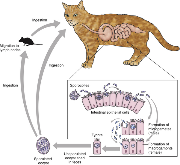
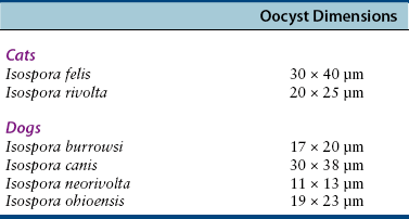
Chapter 82 Isospora spp. are protozoan coccidian parasites that have been recognized for years as potential pathogens in dogs and cats.2–4 The sexual phase of reproduction occurs in the gastrointestinal tracts of dogs and cats and culminates in the passage of oocysts in feces. Cats are the definitive hosts for Isospora felis and Isospora rivolta, and dogs are the definitive hosts for Isospora canis, Isospora ohioensis, Isospora neorivolta, and Isospora burrowsi.2 The oocysts vary in microscopic appearance; this can be used to determine which species is present (Table 82-1).3,4 Isospora spp. are host specific, have worldwide distribution, and infections are very common, particularly in young animals. In the United States, the Companion Animal Parasite Council reports that prevalence rates for Isospora spp. infection in dogs and cats vary from 3% to more than 30%.5 In a study of more than one million fecal specimens from dogs in the United States, 4.4% contained Isospora spp. oocysts.6 In a study of 1355 cats in the United Kingdom, I. felis oocysts were detected in 3%.7 Gender and breed do not usually influence the Isospora spp. shedding rates, but young animals are usually more likely to be shedding oocysts than adults. For example, in one Austrian study, 8.7% of dogs less than 2 years of age were infected; 78% of the positive specimens were in puppies less than 4 months of age.8 In the study from the United Kingdom, I. felis was found in the feces of 9% of cats less than 6 months of age.7 Infection by Isospora spp. in dogs or cats is initiated by ingestion of sporulated oocysts in the environment or by ingestion of tissues of other infected vertebrate “transport” hosts (Figure 82-1).9,10 Infection can also occur if the dog or cat ingests sporulated oocysts carried mechanically by flies, cockroaches, or dung beetles.10 The enteroepithelial phase occurs in the small intestine of infected animals and culminates in the passage of unsporulated oocysts in feces. The prepatent period (time between infection and appearance of oocysts in the feces) and patent period (time that the organism can be detected in the body) vary slightly by the species. In one study of dogs experimentally infected with I. canis, the mean prepatent period was 9.8 days (range, 9 to 11 days, n = 22 dogs), the patent period was 8.9 days (range, 7 to 18 days, n = 20 dogs), and all of the puppies developed diarrhea.9 In contrast, the prepatent period for I. ohioensis in one study was 6 to 7 days, and diarrhea was variable.8 The number of oocysts shed by infected animals can vary dramatically.8,9 Depending on the environmental conditions, sporulation can occur in as little as 12 hours. Clinical disease is most common in young, debilitated, and immunocompromised animals. All the different Isospora spp. replicate in the small intestine, but the regions with the heaviest infection vary by species. FIGURE 82-1 Life cycle of Isospora spp. The host species (in this case, cats) can be infected by ingestion of sporulated oocysts or ingestion of prey that have organisms encysted in their mesenteric lymph nodes. Oocysts shed in the feces can sporulate in as little as 12 hours under optimal environmental conditions.
Isosporiasis
Etiologic Agent and Epidemiology
Clinical Features
![]()
Stay updated, free articles. Join our Telegram channel

Full access? Get Clinical Tree


