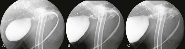
Interventional Oncology

Chapter 76


Interventional Oncology


Although surgery, chemotherapy, and radiation therapy (RT) are being offered in many practices and are the foundations of oncologic treatment, other options are emerging and rapidly gaining acceptance. Interventional radiology (IR) is a specialty that involves the use of image guidance to perform minimally invasive diagnostic and therapeutic procedures. IR techniques are being used more commonly in veterinary medicine to treat neoplastic disease, and this subspecialty of IR, interventional oncology (IO), is expanding. IO provides advanced options to improve the overall standard of care that can be offered to our veterinary patients and is steadily becoming a fourth pillar of oncologic treatment.


Principles


The goal of IO is to perform diagnostic and therapeutic procedures in a minimally invasive fashion. To accomplish this, different imaging modalities are used to allow a clinician to visualize and successfully access a lesion without needing to perform a more invasive procedure. Fluoroscopy is the most readily used imaging modality, and a thorough understanding of the anatomy as revealed by fluoroscopy is crucial to successful performance of IO procedures. Other imaging modalities commonly employed include ultrasonography, computed tomography, and magnetic resonance imaging.


A clinician performing IO procedures needs to have a fully developed understanding of the vast array of available IO instrumentation. Instruments including guidewires, sheaths, diagnostic and guiding catheters, stents, embolic agents, balloons, and various types of contrast are available, and knowing the advantages and disadvantages of the use of each of these items is essential to optimizing the outcome in a particular case.


With the goal of minimizing patient discomfort through the use of minimally invasive techniques, access to organs or regions of the body generally is obtained either percutaneously, through a natural orifice (mouth, nares, anus, or vestibule or penis), or through a blood vessel using the Seldinger technique. In veterinary patients venous approaches do not require vascular repair after the procedure due to the low pressure in the venous system; however, arterial approaches generally do require vessel ligation or repair.


Veterinary Interventional Oncology Treatment Categories


Although IO techniques can be used as supplementary diagnostic tools, IO generally is focused on therapy. The current indications for IO in veterinary medicine fall into three major categories: palliation, primary treatment, and neoadjuvant or adjuvant therapy. Some treatments can overlap across categories, and several different therapies may be offered to an individual patient.


Palliation


Stenting of Malignant Obstructions


Currently IO is used most commonly in veterinary medicine for the palliation of clinical signs associated with neoplastic obstructions. Luminal obstructions secondary to neoplasia occur commonly in veterinary patients and can cause significant morbidity and even mortality. Stents are regularly employed in human medicine for the purpose of opening or “recanalizing” obstructed lumens. The use of stents in veterinary patients is growing rapidly, and several stenting options for the palliative care of animals have been explored.


Urethra.

Urethral stenting has been described in several human studies. Reports of the treatment of malignant urethral obstructions in both dogs and cats have been published (Christensen et al, 2010; Newman et al, 2009; Weisse et al, 2006), and this treatment is now available in many clinics (Figure 76-1). In the first veterinary case series (Weisse et al, 2006), 12 dogs underwent placement of urethral stents to relieve malignant urethral obstructions secondary to prostatic or urethra-bladder neoplasia; in all cases, the dogs were experiencing severe or complete obstruction. All dogs regained the ability to urinate after stent placement. Although urinary incontinence occurred in 25% of cases after stenting, the outcome was considered fair to excellent in 10 of 12 dogs (Weisse et al, 2006). Urethral stenting has been reported in two separate feline case reports of malignant obstruction as well (Christensen et al, 2010; Newman et al, 2009). Both cats were diagnosed with urothelial carcinoma and were noted to have successful urination after stent placement.


Urethral stents are placed through a natural orifice in both dogs and cats. In male dogs, access to the urethra and bladder is straightforward because a guidewire can be introduced easily into the penile urethra. In female dogs and cats, endoscopic or fluoroscopic guidance often is necessary to pass a guidewire into the urethra and bladder. In the authors’ practices, access to the male cat urethra generally requires a surgical approach with a small incision in the bladder and antegrade placement of a stent.


Ureter.

Ureteral obstruction secondary to neoplasia is rare. Primary ureteral tumors that occur cranial to the ureterovesicular junction generally are treated with surgical removal; however, ureteral obstruction secondary to a tumor originating in the bladder or urethra and encompassing the ureterovesicular junction often is not as amendable to resection. Non–urinary tract tumors also can cause extraluminal compression of the ureter. The use of ureteral stents to relieve ureteral obstruction is an important treatment option in humans, and both retrograde and antegrade techniques (using nephrostomy) have been described.


A technique for percutaneous placement of ureteral stents in dogs with malignant ureteral obstruction was reported recently (Berent et al, 2011). In this series of 12 dogs, the obstructed ureters were accessed in antegrade fashion through the placement of a needle within the renal pelvis (successful in 11 of 12 dogs); ultrasonographic guidance was used to position the needle appropriately. In all cases, a double-pigtail stent was placed with the goal of leaving the cranial pigtail in the renal pelvis and the caudal pigtail in the urinary bladder beyond the obstruction. All patients with azotemia demonstrated improvement in blood urea nitrogen and creatinine concentrations after stent placement. Of the 10 patients that underwent abdominal ultrasonography after stent placement, all were demonstrated to have decreased severity of hydronephrosis and hydroureter. Overall, ureteral stent placement was determined to be safe and well tolerated in that cohort of dogs.

< div class='tao-gold-member'>

Only gold members can continue reading. Log In or Register to continue

Stay updated, free articles. Join our Telegram channel

Jul 18, 2016 | Posted by in PHARMACOLOGY, TOXICOLOGY & THERAPEUTICS | Comments Off on Interventional Oncology

Full access? Get Clinical Tree

Get Clinical Tree app for offline access