Inhalation Anesthesia in South American Camelids


Inhalation Anesthesia in South American Camelids


Increased interest in llamas, and more recently in alpacas, as pets and as breeding and pack animals has led to increased demand for veterinary services for them. Although they have some unique species characteristics with regard to anesthesia, many of the principles and techniques used in food animal and equine anesthesia also apply to South American camelids (SACs). Except for differences in size, use of the inhalation anesthetic drugs in alpacas and llamas is similar.


Equipment


Prior to using inhalation anesthesia in SACs, the necessary equipment has to be assembled. Appropriate size endotracheal (ET) tubes and a suitable anesthesia machine are required. Conventional small animal or human ET tubes are appropriate for crias, most juvenile camelids, and some adult alpacas. Larger adult alpacas and adult llamas will require use of ET tubes that are longer and of greater diameter. Commercially available foal endotracheal tubes (50 centimeters [cm] length) are used in those instances. Prior to anesthetic induction verify ET tube length is adequate by placing it alongside the head and neck of the patient.


Conventional small animal anesthesia machines with single sodalime canisters usually do not contain sufficient absorptive capacity for efficient carbon dioxide removal in camelids weighing more than 70 kilograms (kg). Small animal anesthesia machines that have expanded sodalime canisters or dual canisters have sufficient absorptive capacity similar to anesthesia machines designed for use in humans. Anesthesia machines designed for use in adult horses are not recommended for use in camelids.


General Anesthesia


Camelids tend to recover well from anesthesia without experiencing emergence delirium and often are not tranquilized prior to induction unless their temperament necessitates it or use of preoperative sedative or analgesic agents is desired as part of the anesthetic protocol. Camelids are prone to parasympathetic discharge during intubation or with painful stimuli during surgery. Atropine (0.02 milligram per kilogram [mg/kg], intravenously [IV] or 0.04 mg/kg, intramuscularly [IM]) is recommended to prevent bradyarrhythmia and will also decrease salivary secretions. If desired, glycopyrrolate (5–10 microgram per kilogram [mcg/kg], IM; or 2–5 mcg/kg, IV) could be substituted for atropine.


Induction may be accomplished in untranquillized camelids with several different techniques, including thiobarbiturates, if available (8–10 mg/kg, IV); xylazine (0.03–0.05 mg/kg, IV), followed by ketamine (3 mg/kg, IV); diazepam (0.1 mg/kg, IV) combined with ketamine (4 mg/kg, IV); midazolam (0.1 mg/kg, IV) combined with ketamine (4 mg/kg, IV); 5% guaifenesin (50 milligram per milliliter [mg/mL]) and 0.2% thiobarbiturate (2 mg/mL) solution (1.5–2.0 mL/kg, IV to effect); 5% guaifenesin (50 mg/mL) and 0.1% ketamine (1 mg/mL) solution (1.5–2.0 mL/kg, IV, to effect); propofol (4–6 mg/kg, IV), and tiletamine–zolazepam (2.2 mg/kg, IV).


Mask induction with halothane, isoflurane, sevoflurane, or desflurane, as performed in foals or small ruminants, may be used in small or debilitated camelids or to improve muscle relaxation to facilitate intubation in camelids restrained with xylazine–ketamine, tiletamine–zolazepam, and so on. Conventional small animal anesthesia masks may be used, or a mask may be fabricated from a plastic bottle. Sevoflurane and desflurane provide very quick induction of anesthesia and recovery from anesthesia. Currently, halothane is unavailable in the United States, and sevoflurane is a relatively expensive agent to use. Desflurane is less expensive than sevoflurane, but the vaporizer used to administer the agent is very expensive. Economic considerations dictate the use of both drugs in camelid anesthesia, but both offer the distinct advantage of allowing the camelid to regain control of its airway rapidly and thus may help prevent complications associated with airway obstruction following extubation.


Mask induction in healthy, untranquillized adult camelids is usually not attempted because application of the mask may provoke spitting. The technique is usually reserved for camelids weighing less than 50 kg or for obtunded camelids. Oxygen flow rates of 4 to 6 liters per minute (L/min) with vaporizer settings of 3% to 4% halothane; 3% to 4% isoflurane; 5% to 6% sevoflurane; or 10% to 15% desflurane are commonly used. If desired, the mask may be applied and the vaporizer setting gradually increased to allow the animal to acclimate. Addition of nitrous oxide to the gas mixture (50% of total flow) will hasten induction.


Intubation


After induction, tracheal intubation is recommended because it provides a secure airway and prevents aspiration of salivary secretions and gastric fluids if regurgitation occurs. Oral intubation (cuffed tubes, 5–14 millimeters [mm], internal diameter [i.d.]) is performed as in domestic ruminants (Table 47-1). ET tube length and cuff integrity must be confirmed before intubation is attempted. Oral blind intubation is usually unsuccessful, and laryngoscopy, with a 205-mm blade for juvenile and small adult alpacas and a 350-mm blade for adult llamas, is recommended. Because the mouths of herbivores do not open very widely, the blade of the laryngoscope must be placed on the epiglottis to obtain adequate visibility of the larynx for successful intubation. Visibility of the larynx is further improved with the use of a Butler equine mouth gag to open the mouth widely, by hyperextension of the head and neck to make the orotracheal axis approach or exceed 180 degrees, and by use of a gauze sponge on a sponge forceps to swab the pharynx if secretions hinder visibility of the larynx. It is recommended that an obturator, for example, a male canine urinary catheter, be inserted through the ET tube to facilitate intubation.


The obturator should extend 6 to 10 cm beyond the bevel of the ET tube to allow it to be easily visualized as it is passed into the larynx. The ET tube is then threaded off the obturator into the trachea. Attempting intubation when anesthetic depth is insufficient will often provoke active regurgitation when the laryngoscope blade contacts the epiglottis or when the obturator or ET tube contacts the larynx. With adequate depth of anesthesia, this reflex is eliminated. Desensitization of the larynx with topical lidocaine, as is performed in sheep, goats, and swine, is usually not necessary in camelids. However, desensitization of the larynx is helpful when intubation is difficult or anesthesia depth is insufficient.


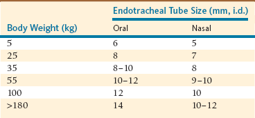
Nasotracheal (NT) intubation in camelids is also possible, although it requires an ET tube one size smaller (Table 47-1). Camelids are prone to epistaxis, so use of lubricating compounds that contain phenylephrine is recommended. Blind nasal intubation is technically easier than oral intubation under laryngoscopic control. Nasal intubation under laryngoscopic control is technically more difficult than orotracheal intubation. Even though NT intubation can be more difficult, it offers the option of recovering the animal with the ET tube in place as a method of preventing airway obstruction during recovery. The ET tube is advanced through the external nares into the ventral meatus with slow gentle pressure, using the same technique used in a horse. If an obstruction is encountered at approximately 6 to 10 cm in adult llamas, it is usually because of placement of the tube in the middle meatus. If an obstruction is encountered more caudally, approximately 25 cm in adult llamas, the tube is likely in the pharyngeal diverticulum. In either case, the tube should be withdrawn and redirected. If the ET tube cannot be redirected past the pharyngeal diverticulum, placement of a prebent stylet (e.g., a piece of the smallest aluminum rod available for Thomas splints) into the tube to direct the ET tube tip ventrally is usually effective. The pharyngeal diverticulum is not as prominent in alpacas.


After the ET tube has been advanced into the nasopharynx, the camelid’s head and neck should be hyperextended and the tube manipulated into the larynx. If the tube will not enter the larynx, placing a prebent stylet in the ET tube to direct the tube tip ventrally into the larynx instead of the esophagus is helpful. Although visibility of the larynx is somewhat limited, oral laryngoscopy will aid intubation and confirm correct or incorrect placement of the tube during intubation.


ET intubation may be confirmed with any of several techniques. Initially, these techniques include visualization of the ET tube passing into the larynx and absence of stertorous breathing sounds. When transparent ET tubes are used, condensation of water vapor will appear and disappear during each breath. Gas can be felt as it is expelled from the tube during exhalation. If a suction bulb (Figure 47-1) is evacuated and connected to the ET tube, it will reexpand if the tube is in the trachea and will remain collapsed if the tube is in the esophagus, thus providing immediate and definitive confirmation of correct or incorrect placement. When the ET tube is connected to an anesthesia machine, observation of synchrony between movement of the rebreathing bag and the thorax will be noted. Finally, if a capnograph or respiratory gas analyzer is available the presence of carbon dioxide (CO2) will be apparent in exhaled gas.


Stay updated, free articles. Join our Telegram channel

Mar 27, 2017 | Posted by in GENERAL | Comments Off on Inhalation Anesthesia in South American Camelids

Full access? Get Clinical Tree

Get Clinical Tree app for offline access