Iatrogenic Damage Caused by Modern Dentistry Procedures

CHAPTER 72 Iatrogenic Damage Caused by Modern Dentistry Procedures


Given the popularity of modern dental procedures, the veterinary practitioner must be aware of the potential damage that can be caused to horses’ teeth. The introduction of modern manual and powered cutting and rasping instruments has led to a substantial increase in the number of instances in which horses’ dentition is damaged.


Often the damage to a horse’s teeth is not detected at the time of the dental procedure, and years may pass before the results of inadvertent damage manifest. The time lapse between the dental procedure and development of clinical signs resulting from the damage may lead to the person performing the dentistry remaining unaware of problems created for the horse. The damage that can be caused is often permanent and extremely painful for the horse but might not be easily diagnosed. The results of the damage can, at the very least, cause a painful pulpitis and behavioral and performance changes and, at the worst, cause death of teeth, abscess formation, loss of teeth, systemic illness, and even death of the horse. A practitioner’s aims should be to do no harm, recognize what is abnormal in the horse’s mouth, and correct abnormalities without causing painful iatrogenic damage to the horse’s teeth.


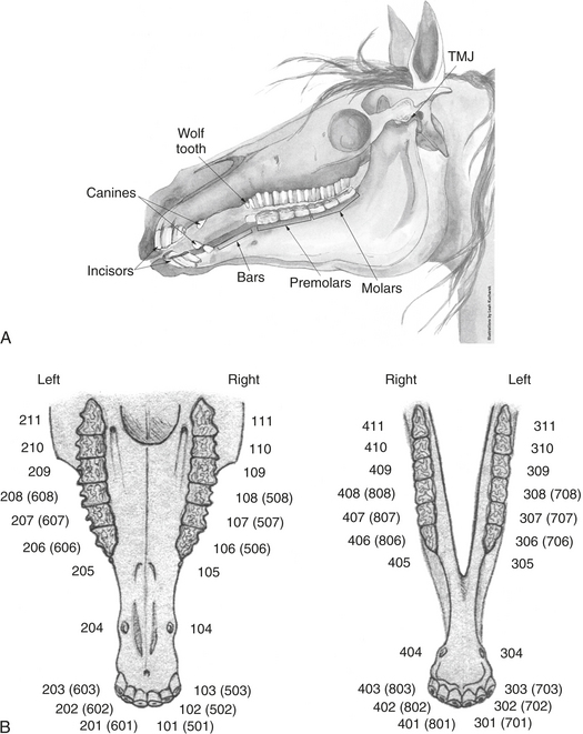
NORMAL DENTAL ANATOMY


It is important to be familiar with normal dental anatomy so that abnormalities can be recognized and corrected without damaging the teeth. In addition, veterinarians should familiarize themselves with the Triadan tooth nomenclature (Figure 72-1). Equine teeth are hypsodont teeth, which have a short erupted crown and a long reserve crown and erupt continuously throughout a horse’s life. The normal structure of equine cheek teeth consists of an intricate pattern of hard enamel folds sandwiched between softer cementum and dentine. The difference in wear between the three materials results in a self-sharpening effect: the sharp enamel becomes exposed as coarse feed materials wear the softer cementum and dentine away. This also leads to raised areas on the occlusal surface in regions where there are an increased number of enamel folds. These lateral-to-medial raised areas, termed transverse ridges, are normal and function to increase the area of the occlusal surface.


Horses have anisognathic head anatomy wherein there is normal disparity in the width of the mandibular and maxillary jaws. This difference in width leads to angulation of the occlusal surface of the teeth, at angles of 10 to 15 degrees. Normal cheek teeth table angles differ greatly among horses as a result of individual conformation. The angulation can also vary between individual teeth or groups of teeth within the same row of cheek teeth. The buccal side of the maxillary cheek teeth and the lingual aspect of the mandibular cheek teeth normally have sharp serrated edges of exposed enamel. If lateral excursion is decreased in the masticatory cycle, sharp enamel overgrowths develop as eruption of the teeth continues. These sharp enamel points cause painful ulcerations to adjacent soft tissues, which further alters mastication. This altered masticatory cycle perpetuates abnormal wear of the cheek teeth and incisors.


The natural curvature of some jaws can make it appear that there is a ramp on the surface of the first lower cheek teeth, and the “curve of Spee” can often give the false impression that there is a hook or high tooth on the last lower mandibular cheek tooth. If these teeth are unnecessarily reduced, iatrogenic malocclusion will be created as the opposing tooth becomes dominant.


Veterinarians should become familiar with the variations in the location and occlusal appearance of the pulp chambers and infundibuli of the maxillary cheek teeth. The maxillary cheek teeth have from five to eight pulp chambers, which can be recognized by the darkly stained secondary dentine. These teeth also have two infundibuli filled with light cream-colored cementum. The mandibular cheek teeth contain only five to seven pulp chambers and no infundibuli. In health, it should be impossible to insert a thin probe into the secondary dentine that seals the pulp chamber, but a probe can often be inserted into a small hole in the center of the cementum of the normal infundibulum. The incisors on a young horse have a single infundibulum (cup) containing cementum.


The incisors also have a pulp chamber that can divide into two or more canals and should be filled completely with dentine (dental star). The middle cheek teeth have five pulp chambers each, and the teeth at the ends of the cheek teeth rows (Triadan system 106, 206, 306, 406, 111, 211, 311, and 411) have additional pulp chambers. Knowledge of the position of these extra pulp chambers is extremely important when overgrowth on these teeth is reduced. The sixth pulp chamber of teeth 106, 206, 306, and 406 is at the rostralmost aspect of these teeth. Additional pulp chambers also exist at the caudalmost aspects of the last cheek teeth.


May 28, 2016 | Posted by in EQUINE MEDICINE | Comments Off on Iatrogenic Damage Caused by Modern Dentistry Procedures

Full access? Get Clinical Tree

Get Clinical Tree app for offline access