Mary Lassaline Utter
Fungal Keratitis
Equine keratomycosis can be a serious disease, resulting in permanent blindness in the affected eye or even loss of the affected globe. Early diagnosis and aggressive management are critical to ensure the best visual and cosmetic outcome and to minimize the length and cost of treatment. Keratomycosis develops when a fungal organism, which may be part of the normal conjunctival flora, inoculates the corneal stroma through a corneal epithelial defect.
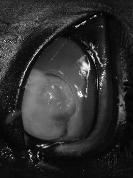
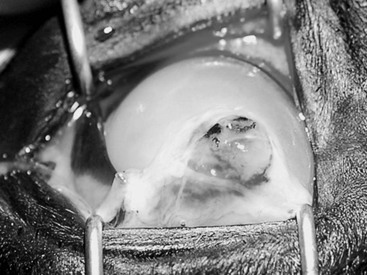
Two clinical presentations include ulcerative keratomycosis, which in severe cases can be associated with stromal melting (Figure 145-1) and globe perforation (Figure 145-2), and fungal stromal abscess (see Color Plate 145-1) in which the epithelial surface of the cornea is intact but there is a fungal infection within the corneal stroma, often as deep as Descemet’s membrane. Although keratomycosis can begin with a superficial stromal infection, both ulcerative keratomycosis and fungal stromal abscess can be associated with deep stromal infection, which can lead to globe perforation in the former case and intraocular abscess rupture in the latter, because fungal organisms have a tropism for the glycosaminoglycans in the deep corneal stroma adjacent to Descemet’s membrane. Neither corneal melting nor corneal stromal abscess requires a fungal infection to develop; both of these diseases can be bacterial or sterile as well as fungus-associated. Both presentations can be associated with profound ocular pain, and both can necessitate extended medical therapy, and often surgical intervention, to achieve resolution.
Keratomycosis can affect any horse and has no breed, age, or sex predisposition. Geographic location is perhaps the most significant risk factor, with the highest incidence found in areas such as the southeastern United States, where fungal organisms are ubiquitous in the environment because of heat and humidity. In the northeastern and mid-Atlantic regions, fungal keratitis is diagnosed more frequently during summer and fall, also associated with heat and humidity. Travel history can provide important information about risk for keratomycosis, particularly for show horses and racehorses that overwinter in southern climates and then move north in the summer.
Horses with a history of prior ocular disease treated with topical antimicrobials or corticosteroids may be at increased risk for developing keratomycosis because these medications can impair the efficacy of the body’s own natural defenses (e.g., antimicrobial substances normally present in the precorneal tear film) as well as alter the resident microbial flora that help modulate growth of opportunistic pathogens.
Diagnosis
Although diagnosis of keratomycosis can be straightforward, with many cases revealing fungal hyphae on corneal cytology or growth of fungal organisms on corneal culture, presumptive early and aggressive intervention based on a high clinical index of suspicion can potentially prevent rapid progression of disease and limit disease severity and duration. Corneal cytology can provide an immediate diagnosis if fungal hyphae are present and in many cases does not necessitate special staining because hyphae are often visible on slides stained only with Diff-Quik, a commercial Romanowsky stain variant. Collecting samples for corneal cytology can be therapeutic as well as diagnostic because the scraping procedure removes fungal organisms as well as unhealthy corneal epithelium and stroma.
Diagnosis can be complicated when fungal organisms proliferate deep in the corneal stroma, or in the face of an intact corneal epithelium that renders the stroma inaccessible to routine corneal sampling techniques for cytology and culture. In cases of fungal stromal abscess, in particular those that resolve without surgical intervention and thus without an opportunity to acquire a deep corneal biopsy for histologic analysis, treatment is necessarily empirical. For cases of stromal abscess in which surgical intervention is elected and a deep keratectomy is performed, excised samples should be submitted for histologic evaluation to obtain a definitive etiologic diagnosis.
The most medically important fungi responsible for equine keratomycosis are the filamentous fungi, which include both Moniliaceae (nonpigmented genera such as Aspergillus and Fusarium spp) and Dematiaceae (pigmented genera such as Curvularia, Alternaria, and Cladosporium spp). Organisms in the former group are most commonly implicated in severe equine keratomycosis. Yeasts (such as Candida spp) are implicated more commonly in human keratomycosis and are not frequent causes of severe equine disease.
Fungal organisms require an injury that penetrates the corneal epithelium, or a previous epithelial defect, to enter the cornea and proliferate. Yeasts are the most common invaders of preexisting epithelial defects, whereas filamentous fungi most commonly invade the corneal stroma in association with trauma, supporting the idea that equine keratomycosis is more often the result of traumatic inoculation than of opportunistic infection by normal ocular microflora.


