21 Feline symmetrical alopecia
CASE HISTORY
As in all dermatology cases, a thorough history (see Chapter 1) is essential in making an accurate diagnosis. Frequently, in cases of symmetrical alopecia, the client may be unaware that the cat is actually overgrooming.
The history in this case was as follows:
• The owner had seen the cat overgrooming the ventral trunk and was aware that the alopecia was self-induced.
• The use of an Elizabethan collar for 3–4 weeks had resulted in regrowth of hair, but the alopecia recurred on removal of the collar.
• There had been a good response to glucocorticoid therapy, with regrowth of hair over the affected area, but treatment resulted in an excessive appetite and weight gain.
• There was one other cat and a dog in the same household. Neither of these animals had developed any skin lesions.
• Fleas had been seen on both cats following a stay in a cattery, prior to the onset of the alopecia. Since then, all animals in the household had been treated with monthly applications of fipronil spot-on and the house environment had been treated with a permethrin and methoprene spray.
• During a 4-week period a commercial, pelleted, chicken hydrolysate diet was fed to both cats, but no change in hair growth was noted during this time.
CLINICAL EXAMINATION
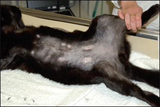
• Examination of the skin revealed non-inflammatory, partial to complete alopecia over the axillae, ventral chest, groin and medial thighs (Fig. 21.1).
CASE WORK-UP
The following diagnostic tests were undertaken:
• Trichographic examination revealed fractured hair shafts from affected areas (see Fig. 2.6, Chapter 2). There was no evidence of ectoparasitism on microscopic examination of scale and skin scrapes from the dorsal trunk, and no evidence of Demodex mites on skin scrapes from the areas of alopecia over the ventrum.
• There was no evidence of dermatophytes on Wood’s lamp examination. No dermatophytes were grown from material collected from the hair coat using the MacKenzie brush technique.
Trial ectoparasitic treatment: Good flea control was already in place, but in order to be confident about ruling out the involvement of cheyletiellosis, all animals were treated with fortnightly applications of selamectin for a period of 4 weeks. There was no response to this and the cat continued to overgroom. Monthly applications of fipronil were continued after the trial selamectin therapy.
Stay updated, free articles. Join our Telegram channel

Full access? Get Clinical Tree


