Feline glaucoma

46 Feline glaucoma


CLINICAL EXAMINATION


General clinical examination is frequently unremarkable. One or both eyes can be affected. The pupil is normally dilated and the direct pupillary light reflex absent or sluggish and incomplete (Figure 46.1). Moderate episcleral congestion is usually present but this can be difficult to appreciate since the globe fits tightly into the socket and there is little exposed sclera or episcleral tissue. Sometimes chemosis may be noted. Corneal oedema is not a common feature of feline glaucoma.


Globe enlargement can be present but is often subtle and needs to be differentiated from exophthalmos. Looking down on the eyes from above will help since retropulsion is minimal in cats and thus can be difficult to appreciate, while looking down from above one can visualize if the globe fits in the socket or is pushed forwards while also comparing the corneal curvature of the two eyes – this will be altered if the globe is truly enlarged. Sometimes the globe enlargement will lead to an exposure keratitis – corneal ulceration and vascularization may be present and fluorescein testing should always be undertaken.


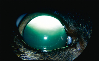
Signs of chronic uveitis might be present, and these could include keratic precipitates, rubeosis iridis, iris lymphoid nodules, posterior synechiae and iris rests (Figure 46.2).


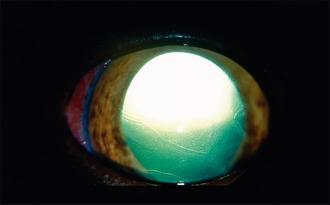
The lens can be cataractous and is sometimes luxated into the anterior chamber. Another dissimilarity with dogs is that the luxated lens does not tend to further exacerbate the glaucoma and develops as a result of the uveitis damaging the lens zonules, not due to primary lens instability. Lens luxation in cats is less painful than in dogs since the anterior chamber can accommodate the dislocated lens without further exacerbating the glaucoma.


If possible, fundus examination should be performed and this will frequently reveal diffuse tapetal hyper-reflectivity and blood vessel attenuation as signs of chronic retinal degeneration. The optic disc will be cupped but this can be difficult for general practitioners to appreciate since the feline disc is small and dark anyway, and the signs of feline disc cupping are much more subtle than in dogs.


Intraocular pressure measurement is essential and access to tonometry should be made available. Cats can have dramatically high intraocular pressures – in the 60s mmHg sometimes – yet show remarkably little pain and some will even maintain some useful vision despite the very high readings. Always compare the readings in the two eyes since the difference between them can help to diagnose borderline cases. For example, a cat with an intraocular pressure of 28 mmHg in one eye but 8 mmHg in the other, where both eyes have signs of uveitis, would certainly have secondary glaucoma, while a cat with readings of 26 mmHg in both eyes, and no other ocular changes, might simply have high normal pressures.


If there are no signs of previous or active uveitis, then assessing the depth of the anterior chamber can be very useful. A rare form of feline glaucoma manifests with a shallow anterior chamber following misdirection of aqueous into the vitreous gel, forcing the iris and lens more anterior. The drainage angle can be easily assessed in cats without the need for goniolenses – simply shining a pen torch across the cornea and looking at the angle between the iris and cornea will reveal the pectinate ligaments as the opening to the uveoscleral drainage angle. Goniodysgenesis is not a feature in feline glaucoma, but the angle can be narrowed if the iris is pushed forwards, and is frequently blocked by inflammatory cells or peripheral anterior synechiae following anterior uveitis.


CASE WORK-UP


If the glaucoma is secondary to anterior uveitis, as is most common in cats, then the cause for the uveitis should be investigated and this is discussed in Chapter 34. If a uveal melanoma is suspected, the cat should be thoroughly assessed for signs of metastasis with blood work, radiography and ultrasonography. If there is no evidence of other intraocular disease, then a tentative diagnosis of primary glaucoma is made. It has been seen in Burmese and elderly Siamese cats on occasion, and the rare aqueous misdirection form mentioned briefly above can be seen in any cat breed. If a primary glaucoma is suspected, then referral should be considered, particularly to confirm this diagnosis and provide prognostic information for both eyes.


Stay updated, free articles. Join our Telegram channel

Sep 10, 2016 | Posted by in SMALL ANIMAL | Comments Off on Feline glaucoma

Full access? Get Clinical Tree

Get Clinical Tree app for offline access