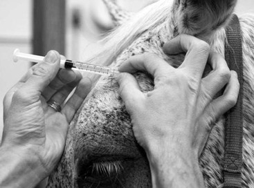
Caryn E. Plummer Most ocular structures can be observed either directly or indirectly, and a thorough ophthalmic examination can lead to a rapid and accurate diagnosis for many ophthalmic diseases. The complete ophthalmic examination includes obtaining historical data, observing the animal’s behavior and reactions in both well-lit and dim-light conditions, examining the ocular structures in a darkened environment, and collection of diagnostic or supportive data and samples as indicated. The eye lends itself to numerous simple and efficient diagnostic procedures, many of which can be performed during a routine examination. Most of these procedures are noninvasive, and a thorough understanding of them can facilitate identification, diagnosis, and the forming of a prognosis. A thorough ocular and medical history should be obtained and should include the horse’s signalment, its intended use, and descriptions of its home environment, as well as specific information related to the reason for its presentation. The handler should be questioned about the initial clinical signs and their onset, progression and duration of the problem, previous and current treatments given, response to those treatments, and any concurrent nonocular signs or behavioral changes. A general physical examination should accompany a thorough ophthalmic examination, especially if the ophthalmic condition is accompanied by systemic signs. Also, because of the close relationship of the eye to the central nervous system, a complete neurologic examination may be indicated in horses suspected of having neuroophthalmic disease. The initial examination of the eye should take place before sedation and should be performed from a distance. The animal’s overall symmetry and body condition, attitude, and ability to navigate are carefully evaluated. The horse should be watched closely as it is led to the examination area, especially if it is an unfamiliar environment. Balking at objects or obstacles or hesitation when turning or being led in another direction may provide some insight into the state of the animal’s vision. The horse’s comfort and symmetry should be assessed by observing for blepharospasm, globe position and periocular symmetry observed from both the front and the side, and eyelash symmetry. Deviation of the lashes downward can be a subtle indication of ocular pain, enophthalmos, or lid dysfunction (ptosis, facial nerve paralysis). The menace response in each eye, as well as other subjective assessments of vision (e.g., maze testing alternately covering eyes with blinkers), are then tested. If the horse has sight, a quick, threatening gesture or movement of the hand toward the eye should result in a blink or movement of the head away from the stimulus. Care should be taken not to create air currents toward the eye when performing this test, which would test sensation rather than vision. The menace response is an acquired response and is therefore not exhibited by some normal young animals, especially foals younger than 2 weeks of age. Pupillary light, dazzle, and palpebral reflexes are also tested before the administration of any pharmacologic agents. The pupillary light reflex evaluates the integrity of the retina, optic nerve, midbrain, oculomotor nerve, and iris sphincter muscle. The normal equine pupil may respond somewhat sluggishly and incompletely unless the stimulating light is especially bright. This stimulation of one eye should result in the constriction of both pupils; that is, there should be a consensual response in the fellow eye. Shining a light in the fellow eye and observing this consensual response is a valuable test of retinal function in eyes in which the direct pupillary light reflex is not appreciable because of severe corneal or anterior chamber opacity or previous atropine administration, for example. Blinking or squinting is the normal response to shining a very bright light into one eye. This dazzle reflex is subcortical and requires functioning of the retina, optic nerve, and facial nerve. A bright, focal light source must be used, particularly in animals with lesions obstructing the visual axis. The palpebral reflex should be tested in horses that fail to blink completely, for example, when the menace response is being evaluated. The medial and lateral canthi are gently touched, which normally will stimulate a reflex blink. The basic equipment necessary to perform a proper ocular examination is summarized (Box 140-1). It should include a bright, focal light source such as a Finnoff transilluminator. It may be helpful to develop a recording system for your observations. Written descriptions as well as drawings are minimal requirements for thorough medical record keeping. These techniques may be augmented by digital photography. With practice and patience, good-quality ocular images can be achieved with an inexpensive camera system with 4 megapixels or greater. Archiving the images in the horse’s medical record and photographing the eye at later intervals help determine progress of cases. Most horses need sedation and regional motor blocks to enable thorough examination. The degree of restraint depends on the animal’s temperament, response to tranquilizers, and degree of discomfort. Intravenous sedation with an α2 agonist is usually sufficient for an ophthalmic examination. Additional sedation or the addition of an opioid such as butorphanol may be necessary for more painful or invasive procedures. Auriculopalpebral blocks facilitate examination of the equine eye. This motor block results in akinesia of the palpebral branch of the facial nerve, which innervates the orbicularis oculi muscle that is responsible for eyelid closure. Because the equine eyelid is very strong, this motor block is critical if the eye is painful or if the integrity of the globe is compromised such that the struggle required to open the upper eyelid without the block might result in globe rupture. For this block, 1 to 2 mL of an anesthetic (lidocaine, mepivacaine, bupivacaine) is injected subcutaneously through a small-gauge needle (25 gauge) laid adjacent to the nerve. Care should be taken to stabilize the needle hub during injection so that the needle does not break off the hub into the skin. If there is resistance during injection, the needle should be redirected because the tip is likely intradermal rather than subcutaneous. The palpebral branch of the auriculopalpebral nerve can be palpated and blocked just lateral to the highest point of the caudal zygomatic arch or where it lies on the zygomatic arch caudal to the bony process of the frontal bone (Figure 140-1). The auriculopalpebral nerve can be blocked closer to its origin in the depression just anterior and ventral to the base of the ear where the caudal border of the mandible meets the zygomatic process of the temporal bone. Paralysis lasts 1 to 2 hours. Horses that have received repeated auriculopalpebral nerve blocks occasionally become resistant to the effects of the anesthetic, which manifests as a longer time needed for onset of akinesia or a higher dose of anesthetic needed to achieve this effect. If anesthesia of the upper lid is required, such as when an eyelid laceration is to be repaired or a subpalpebral lavage treatment system is to be installed, a frontal (supraorbital) nerve block is indicated. The frontal nerve, which provides sensation to the medial and central upper eyelid, may be blocked as it exits the supraorbital foramen within the frontal bone. This depression can be located by placing one’s thumb below the dorsal orbital rim and the middle finger above the dorsal orbital rim (within the supraorbital fossa). When the index finger is placed between the other two fingers, it should land in the general vicinity of the foramen. Again, as with the motor block, 1 to 2 mL of a local anesthetic agent is injected subcutaneously. If anesthesia of the lateral upper eyelid or lower eyelid is required, line blocks along the edge of the dorsolateral and ventral orbital rims, respectively, will be necessary. A thorough examination of the ocular surface, and if possible, the interior of the globe, permits a quick assessment for ocular disease; however, a precise diagnosis and development of an appropriate course of therapy often necessitate input from additional diagnostic tests. Certain tests or observations must precede others to avoid interference or complications (Box 140-2). Microbiologic samples must be collected before instillation of any diagnostic drugs. If keratoconjunctivitis sicca is suspected, the Schirmer tear test should be performed before excessive ocular manipulation and before instillation of any ophthalmic solutions or ointments. For example, application of topical anesthetic agents will result in falsely low test values. Tonometry should be performed before the instillation of mydriatic agents to avoid inaccurate estimates or elevations of intraocular pressure that may further damage a glaucomatous eye.
Examination of the Eye
Initial Examination
Sedation and Regional Nerve Blocks
Diagnostic Testing
![]()
Stay updated, free articles. Join our Telegram channel

Full access? Get Clinical Tree


Examination of the Eye
Chapter 140
