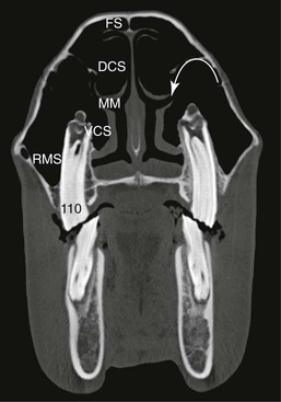
Thomas H. Witte The rostral extent of the nasal cavities, the external nares, is supported by cartilage and muscle. This makes them mobile and flexible, allowing for sealing of the nasal cavity during swimming and widening at maximal exercise, but also allowing for pathologic collapse when there is neuromuscular dysfunction. The clinically significant landmarks of this region include the nasolacrimal ostium at the ventral aspect at the mucocutaneous junction, which is easily cannulated there, and the nasal diverticula, or false nostrils. These blind-ending, hair-lined pockets are closely associated with the lateral aspect of the external nares, from which they are separated by the alar cartilages. Continuing caudally, the midline cartilaginous nasal septum divides the nasal cavity into left and right halves. These cavities are further subdivided by the dorsal and ventral turbinates into the dorsal, middle, ventral, and common meatus (Figure 50-1). The turbinates are scrolled bones that project into the nasal cavity from the lateral wall. The internal space within each scroll is divided into rostral and caudal cavities. The rostral components are termed recesses and are less clinically significant than the caudal components, which form the dorsal and ventral conchal sinuses and communicate with the frontal and rostral maxillary sinuses, respectively. The submucosa of the nasal cavity is highly vascular, in keeping with the function of warming and humidifying inspired air. Because the nasal cavity is encased in rigid bone, any space-occupying lesion here will have a marked impact on airflow and hence athletic performance. Simple vascular congestion caused by iatrogenic jugular thrombophlebitis or loss of sympathetic tone in Horner’s syndrome can be performance limiting and, if left undetected, will limit the success of other upper respiratory tract interventions. The caudal extent of the nasal cavity is bounded dorsally by the middle turbinate, which is surrounded on its caudal aspect by the fine, highly vascular cartilaginous ethmoturbinate scrolls, collectively termed the ethmoidal labyrinth. The increased surface area provided by the labyrinth is lined with olfactory epithelium and with sensory neurons whose axons converge to form the olfactory nerve (cranial nerve [CN] I). Although the integrity of the cribriform plate caudal to the ethmoid is crucial when considering treatment options in this area, it cannot be examined endoscopically in the normal horse. The paranasal sinuses lie lateral and dorsal to the nasal cavity, and although they are made up of six paired chambers, they can be considered as two functional compartments: rostral and caudal. The two compartments drain separately to the middle meatus through the rostral and caudal maxillary sinuses and a common aperture. The nasomaxillary aperture cannot be seen directly with standard endoscopic techniques and equipment, and under normal circumstances, the paranasal sinuses cannot be accessed directly through the nose. Endoscopic evidence of sinus disease is therefore often limited to fluid egress at the drainage angle of dorsal and ventral turbinates in the dorsal nasopharynx, rostral to the middle turbinate. The caudal paranasal compartment consists of frontal, dorsal conchal, caudal maxillary, and sphenopalatine sinuses. The rostral compartment, which is separated from the caudal compartment by the oblique, curved maxillary septum and its dorsal continuation as the bulla of the ventral conchal sinus, consists of rostral maxillary and ventral conchal sinuses. The rostral maxillary and ventral conchal sinuses communicate over the infraorbital canal and are separated ventrally by the septum that supports this structure, or the maxillary cheek teeth, depending on the age of the horse (see Figure 50-1). The caudal outpouching of the ventral conchal sinus is known as the bulla of the ventral conchal sinus and represents a key surgical landmark. The channel for drainage from both the caudal and rostral sinus compartments into the middle meatus is closely associated with this structure. The conjoined sphenoidal and palatine sinuses (known as the sphenopalatine sinus) extend under the ethmoid and the base of the skull (see Color Plate 50-1). Any space-occupying lesion in this area can cause compression of the optic nerves, brain, and pituitary gland, resulting in unusual presenting complaints. Health of the paranasal sinuses in any species relies on adequate ventilation through the natural ostia and on mucociliary transport provided by ciliated columnar epithelium. The mucociliary escalator works toward the natural drainage channels from the rostral and caudal maxillary sinuses, maintaining a continuous protective mucus layer flowing out of the sinuses. This flow is aided by gravity when the head is down, but continues against gravity at other times. The narrow drainage channels are easily obstructed and distorted by primary disease processes, and the resultant impaired drainage must be recognized and addressed. Surgical interventions should be planned carefully with the goal of maintaining or restoring normal mucociliary clearance from the dependent sinuses, rather than creating new drainage ostia in a separate location. Termed functional sinus surgery in humans, this approach aims to be mucosa sparing when possible. The evolution of endoscopic techniques has contributed to the revolution in surgery of the paranasal sinuses. The relationship between the cheek teeth and the paranasal sinuses is clinically significant. Part of the upper Triadan 08 tooth and the entire root system of the 09 are usually associated with the floor of the rostral maxillary sinus, whereas the roots of the 10 and 11 are associated with the floor of the caudal maxillary sinus (see Color Plate 50-1). Periapical disease of these teeth commonly results in secondary sinusitis, compared with periapical disease of the more rostral teeth (06 to 08), in which facial swelling or external draining tracts are more common. The proximity of the teeth makes trephination of the maxillary sinuses difficult and often unrewarding, particularly in the young horse when the reserve crowns are large and the sinus cavities small. The precise relationship between the apices of the cheek teeth and the respective sinuses cannot be predicted on the basis of plain radiographs: disease of the 08 and 09 teeth can result in sinusitis of the caudal compartment if there is rostral extension of the caudal maxillary sinus and obliquity of the maxillary septum, whereas apical disease of the 10 and 11 teeth can result in sinusitis of the rostral compartments when there is caudal extension of the ventral conchal sinus. Attempting to predict which tooth is infected based on which sinuses are diseased should be avoided. Horses with sinonasal disease can have clinical signs ranging from subtle poor performance, epiphora, or head shaking to the more common nasal discharge, facial swelling, and difficult breathing. Because many horses are not presented until the signs are obvious, disease can be at an advanced stage by the time the horse is first evaluated. Understanding the progression historically can be vital to establishing a valid differential diagnosis list and ensuring that appropriate diagnostic tests are undertaken. In particular, changes in the volume, odor, timing, nature, duration, and laterality of discharge over time can be extremely informative. The head should be evaluated from both sides and in front for symmetry and swellings as well as for pain on palpation. The nares should be dry and free from crusting, except for a small volume of tears evident at the nasolacrimal ostium. Airflow should be tested at the external nares; subtle differences may be more obvious after temporary occlusion of airflow or by the use of strands of cotton to detect airflow. Horses with restricted airflow at any level of the respiratory tract may dilate their nostrils bilaterally. The nostrils should remain mobile and tactile and respond to internal and external palpation. Deformation of the facial contour over the paranasal sinuses indicates pressure buildup within the sinuses and most commonly points to a space-occupying lesion such as a cyst or neoplasia. Severe secondary sinusitis cannot, however, be ruled out as a cause of facial distortion because complete occlusion of drainage can result in sufficient pressure to cause distortion. Although percussion of the sinuses for fluid- or soft tissue–induced dullness and palpation of the lymph nodes should form part of any routine physical examination of the head, the findings are often equivocal and difficult to interpret definitively. Endoscopic examination for nasal and paranasal sinus disease may include rhinoscopy, dentoscopy, and sinoscopy. Rhinoscopy can be performed with a standard flexible endoscope. A videoendoscope allows documentation of findings for serial evaluations and can be particularly helpful in treatment of mass lesions. In the presence of active discharge from the nose, rhinoscopy will confirm the source of the drainage. Routine rhinoscopy should include careful evaluation of the nasopharynx, ethmoidal labyrinth, drainage angle between dorsal and ventral turbinates, and mucosa of the dorsal, ventral, and middle meatus. Dentoscopy or examination of the dental occlusal surfaces with a mirror should be undertaken in any horse with unilateral nasal discharge of unknown etiology and must involve a thorough assessment of the periodontium. Careful documentation of findings enables serial monitoring and helps in client communication. Sinoscopy has become less critical as a diagnostic modality for paranasal sinus disease with the advent and availability of standing computed tomography (CT), although it remains a key tool therapeutically and a cornerstone of the diagnostic process if CT is unavailable. Several approaches to each of the sinus compartments have been described, but the most universal appears to be creation of a frontal sinus portal of an appropriate size to allow a flexible endoscope to be inserted, with or without space to pass an instrument alongside depending on surgeon preference (see Color Plate 50-1). Primary approaches to the maxillary sinuses pose the risk for iatrogenic damage to the cheek teeth and are less useful in gleaning an overview of the global disease state, although they may still be very useful therapeutically when they can be placed under sinoscopic guidance or with the benefit of CT. Sinoscopy is performed in the standing sedated horse, making orientation easier and preventing loss of visibility in the presence of bleeding. Suction should be available if possible. A Galt trephine is used to cut a circular disk of bone from the bone plate. Frequent lavage of the site will prevent the buildup of bone dust subcutaneously and minimize postoperative reaction. A 2.5-cm-diameter trephine appears to be the optimal size because this allows an instrument to be passed alongside the endoscope, avoiding the necessity for a second portal, while not being too large for a good cosmetic outcome. Creating two separate 1-cm trephine holes for the endoscope and instrument is also suitable. The frontonasal suture line should be avoided when possible (see Color Plate 50-2). In general, this suture line runs between the medial canthi of the eyes, and placing a trephine or other osteotomy across this line, which becomes more likely with increasing osteotomy size, results in more or less severe suture periostitis. The standard approach to the frontal sinus, caudal to and midway along a line drawn perpendicular to the midline at the level of the medial canthus of the eye, allows the caudal maxillary and conchofrontal sinuses to be explored directly without risk for damage to the intrasinus component of the ethmoid during the approach. Damage to the latter results in profuse hemorrhage, making accurate diagnosis and intervention more difficult. From the frontal sinus, the endoscope can be passed freely through the large frontomaxillary aperture into the caudal maxillary sinus (see Color Plate 50-1). The ventral conchal bulla, the caudal outpouching of the ventral conchal sinus, lies beneath the rostral margin of the frontomaxillary aperture. This cartilaginous bulla, which varies among horses in shape and size and can appear to be absent in horses with chronic deformation of the intrasinus anatomy, can be resected to give access to the rostral maxillary and ventral conchal sinuses. The ventral conchal sinus is often the site of mucus inspissation in cases of chronic sinusitis, and this condition can be confirmed and treated by this approach. When necessary, the frontal portal can be supplemented with a maxillary trephine, which can safely be created under endoscopic guidance and transillumination. Postoperative lavage tubes can be placed through the frontal sinus portal, allowing adequate lavage without creating any further trephines. The pressure of postoperative lavage fluid must be carefully controlled to avoid subcutaneous accumulation of fluid, especially with larger osteotomies. Standard lateral and oblique radiographic views of the paranasal sinuses provide a useful baseline, although more specialized views, including intraoral and open-mouth radiographs, are necessary to gain a more complete understanding of the underlying pathology. A dorsoventral view is particularly useful to identify opacification of the ventral conchal sinus or deviation of the nasal septum. Internal dental architecture can also be visualized if the mandibular and maxillary arcades can be offset. Numerous other special projections have been described for imaging the cheek teeth without superimposition and are based on an understanding of the curvature of the dental arcade and the caudal-to-rostral narrowing evident when one carefully examines the equine skull. The ideal projection to isolate the maxillary cheek teeth is thus taken 15 degrees caudal to the straight lateral and 30 degrees dorsal to the horizontal axis. Nuclear scintigraphy can be helpful in determining the clinical significance of a radiographic finding or, more often, can create a suspicion of dental or sinus origin for spurious or vague presenting signs. Lateral and dorsoventral scintigraphic views of the head are easy to acquire and, depending on signalment and history, may form part of any full-body bone scan performed for poor performance. Ultimately, in difficult, refractory, or recurrent cases, referral for CT may be considered necessary, in particular where this modality is available for the standing horse. This technique is considered the gold standard for complete evaluation of intrasinus space-occupying lesions, particularly when neoplasia is suspected, and for the definitive detection of dental pathology. Computed tomography offers advantages in surgical planning, enabling the examiner to identify the relationship between diseased areas and important structures such as the infraorbital nerve, nasolacrimal duct, and cribriform plate. This information allows the application of advanced, minimally invasive, and functional endoscopic techniques for the treatment of the full range of sinus diseases. The term atheroma is sometimes inappropriately used to describe the variably sized soft, fluctuant swellings found within the nasal diverticulum in horses around the age of 2 years. These swellings are epidermal inclusion cysts that enlarge continuously from exfoliation of squamous cells from their lining. Most commonly these lesions do not threaten the lumen of the airway because they bulge laterally, and their significance is rarely anything more than cosmetic. Diagnosis is based on the pathognomonic location and gross appearance of the lesion, and aspiration yields a gray, odorless, greasy fluid. The masses can be removed intact through an external skin incision, and injection of formalin has been reported, but the most predictable cosmetic outcome is achieved by incising and removing the cyst lining with a ventriculectomy burr placed through the external nares. Alar fold collapse causes an abnormal upper respiratory tract noise that must be differentiated from laryngeal and pharyngeal dysfunction. The noise may be heard at rest or may only be elicited by intense exercise, and has variously been described as a muffled rattling or vibrating noise heard during both inspiration and expiration. Horses may be presented for evaluation primarily because of the noise or because of poor performance. Dynamic respiratory endoscopy can be useful in ruling out other causes of abnormal upper respiratory tract noise and poor performance, but definitive diagnosis necessitates temporary retraction of the alar folds with sutures. By use of a twitch and local anesthesia, suture material is placed through each alar fold and tied dorsally over the nasal bones, occluding the nasal diverticula. Absence of noise after suture placement and recurrence with suture removal provides a definitive diagnosis and justifies surgical resection of the alar folds. Surgery can be performed with the horse anesthetized and positioned in dorsal or lateral recumbency, or with the horse standing. The procedure can be performed through an intact nostril, but some surgeons prefer to incise the lateral alae of the nostrils to expose the alar folds more completely and to ensure complete resection of all associated tissue.
Diseases of the Nasal Cavity and Paranasal Sinuses
Clinically Relevant Anatomy and Physiology
Examination
Historical Findings
Physical Examination
Endoscopy
Diagnostic Imaging
Diseases of the Nasal Cavity
Epidermal Inclusion Cysts
Alar Fold Collapse
![]()
Stay updated, free articles. Join our Telegram channel

Full access? Get Clinical Tree


