Dermatophytosis in a guinea-pig

10 Dermatophytosis in a guinea-pig


CLINICAL EXAMINATION


A careful physical examination should always be carried out to evaluate for any other diseases or additional problems. This guinea-pig was bright, alert and vocal on presentation, and general physical examination was unremarkable, other than the dermatological abnormalities.


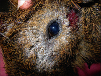
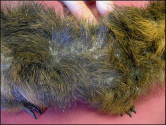
Dermatological examination showed marked crusting and scaling around the left eye (Fig. 10.1), with evidence of inflammation of the skin below the left ear. There was also extension of patchy alopecia and scaling onto the dorsum (Fig. 10.2).


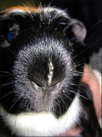
The clinical signs associated with dermatophytosis in guinea-pigs vary. Lesions often begin with circumscribed or coalescing oval to patchy areas of non-pruritic scaling and alopecia, usually affecting the nose (Fig. 10.3), ears and face. However, in advanced cases lesions may spread to involve the neck, limbs and body. If untreated, then the lesions can become inflamed and infected with bacteria. This secondary infection will manifest as pustules, papules and crusts, usually with an increase in the pruritus, although the level of irritation does differ between individuals. Dermatophytosis can occur concurrently with Trixacaris caviae infestation; in these cases, the pruritus tends to be severe.


CASE WORK-UP


Because of the history and the nature of the lesions, dermatophytosis was suspected, although ectoparasitic infestations needed to be ruled out.


Skin/hair scrapes: Multiple skin scrapes were taken from around the left eye and the dorsum. These were mounted in liquid paraffin on a microscope slide with a coverslip and examined under ×40 magnification. No ectoparasites were seen. This absence does not entirely rule out ectoparasites and, in general, it may be useful to repeat the tape strips, or even consider prophylactic antiparasite treatment.


Further investigations aimed towards diagnosing dermatophytosis:


Wood’s lamp examination: This was performed and was negative. Wood’s lamp is an ultraviolet light that should be allowed to warm up for 5–10 minutes prior to use because the stability of the light’s wavelength and intensity is temperature dependent. When exposed to ultraviolet light, hairs invaded by M. canis will show apple-green fluorescence in approximately 50% of isolates. The fluorescence is due to tryptophan metabolites produced by the fungus and hairs should be exposed for 3–5 minutes. Fluorescence is not present in scales or crusts, and illumination of debris should not be mistaken for fluorescent hairs. Trichophyton spp. will not fluoresce under ultraviolet light, therefore Wood’s lamp examination is not often helpful in guinea-pigs and a negative result does not exclude dermatophytosis.


Hair plucks: Examination of hairs from the affected areas may demonstrate infected hair shafts. This is a difficult technique, requiring experience and a high-quality microscope. The hairs should be mounted in liquid paraffin or potassium hydroxide, covered with a coverslip and examined under ×400 magnification. Fungal hyphae may be seen, or arthrospores may be apparent within infected hair shafts. No fungal elements were seen in this case.


Skin/hair scrapes: Scrapes taken from the periphery of lesions can be examined as above for infected hair shafts. In rare cases of Trichophyton spp. infection, hyphae may only be present in the stratum corneum.


Fungal culture: This test enabled definitive diagnosis and identification of the dermatophyte species. Hair pluck and skin scrapes can be used from the lesions. The differences and limitations of in-house versus external testing are described below. In this case, samples were taken for external laboratory fungal culture, and 2 weeks later the results were reported as positive for T. mentagrophytes.


In-house testing: In-house dermatophyte test medium (DTM) kits are available and often used. These comprise a small culture plate containing Sabouraud’s dextrose agar with cyclohexamide, gentamicin and chlortetracycline as antifungal and antibacterial agents. They also contain a phenol red colour indicator. Growing dermatophytes initially use proteins, producing alkaline metabolites that turn the indicator from yellow to red. Often, contaminant fungi use carbohydrates first, producing acidic metabolites which leave the indicator yellow. However, when the carbohydrates are used up, protein metabolism then causes the red colour change. Black or grey colonies are not clinically significant, whatever the colour change.


Hairs (and scale) from the lesion should be collected and inoculated onto the agar, with the lid replaced loosely to allow entry of air. Some dermatophytes take longer than others to produce the red colour change (10–14 days), so the test kit needs to be checked daily for 10 days. In addition, some non-pathogenic dermatophytes will cause the red colour change (e.g. Aspergillus spp.).


Since the presence of fungal growth and colour change is not entirely reliable, microscopic examination is essential to prevent an incorrect presumptive diagnosis.


Once grown, the plate should be sent to an external laboratory for identification. This allows the clinician to be sure what species is causing the problem, and may suggest the source of infection. For example, if M. canis was identified you might consider sampling the house cat.


External laboratory testing: Commercial laboratories use similar culture media but without the colour indicator. Some of the additives to the Sabouraud dextrose agar may inhibit the growth of certain pathogens; therefore, laboratories use plain agar, which sometimes makes the diagnosis of dermatophytosis easier. In addition, the greater experience of laboratory technicians at interpreting culture results and the possibility of extended culture under controlled conditions if required enables more accurate identification of the dermatophyte. Even with in-house DTM kits, the dermatophyte should still be sent to a lab for identification, so sending to the lab in the first place may well save time and be more accurate.


The relative costs of in-house DTM kits and external laboratory fungal culture tend to be fairly similar.


Sterile toothbrush: Samples may be obtained for microscopy or culture by grooming the hairs on and around the lesions with a sterile or new toothbrush.


Skin biopsy: Diagnosis can be made with skin biopsies submitted for histopathology. Fungal elements may be demonstrated with PAS (periodic acid–Schiff) or methenamine stains. Biopsies were not required for diagnosis in this case.

Stay updated, free articles. Join our Telegram channel

Sep 3, 2016 | Posted by in SMALL ANIMAL | Comments Off on Dermatophytosis in a guinea-pig

Full access? Get Clinical Tree

Get Clinical Tree app for offline access