Chapter 79: Tear Film Disorders of Cats

Web Chapter 79


Tear Film Disorders of Cats


Cats with chronic keratoconjunctivitis and, to a lesser degree, blepharitis are encountered commonly in small animal practice. Qualitative tear film abnormalities often are overlooked in the feline patient but can play an important role as either a cause or an outcome of these disorders. This chapter reviews the structure and function of the mucin and lipid components of the precorneal tear film (PTF), highlights their importance in surface ocular disease, and discusses treatment strategies to address tear film dysfunction.


Anatomy and Physiology


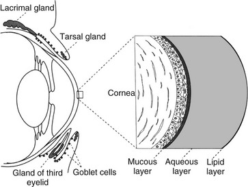
The PTF is a mixture of lipid, aqueous, and mucin components, with regional variation in the concentration of these components (Web Figure 79-1). The exact thickness of the feline PTF has yet to be established; past measurements of the PTF have estimated its thickness to be 7 to 10 µm, with aqueous layer being most abundant. However, research into the human PTF has yielded variable results, including the suggestion of a much thicker tear film consisting mainly of mucin.


The lipid layer is composed of a mixture of polar and nonpolar lipids that tends to be more concentrated at the outermost portion of the PTF. The feline lipid layer resembles that of humans, being thinner than that in dogs. The meibomian or tarsal glands, located within the eyelids, are the main source of PTF lipids. Tear film lipid, or meibum, exists as a liquid at body temperature, which allows it to be spread across the surface of the PTF with eyelid movement. This layer minimizes evaporation of the PTF between blinks and decreases the surface tension of the tear film, thereby maintaining tear film stability and a smooth, optically clear surface.


The mucin component is concentrated at the innermost aspect of the PTF. The majority of ocular mucins are secreted by the conjunctival goblet cells, with lesser contributions from the epithelial cells of the conjunctiva and cornea as well as the orbital lacrimal gland. Although goblet cells are located throughout the conjunctiva, they are particularly concentrated within the ventromedial conjunctival fornices. Mucin plays an integral role in maintaining tear film stability by decreasing the surface tension of the tear film; it also binds contaminants such as particulate matter and bacteria, and aids in maintaining an optically smooth surface by filling irregularities of the corneal epithelial surface. Because of its viscous nature, mucin also helps the hydrophilic tear film adhere to the hydrophobic corneal epithelium.


Although tears are responsible for both conjunctival and corneal surface health, the cornea’s lack of blood supply renders the effects of tear deficiency more notable there than on the conjunctiva; nevertheless, conjunctival effects also remain important. Any alteration of either the lipid or mucin layer is referred to as a qualitative tear film abnormality, whereas a deficiency of the aqueous component is termed a quantitative tear film abnormality.


Pathophysiology


Because of contributions from the lipid and mucin layers, the PTF maintains itself as a continuous film over the cornea. During each blink, fresh tears are spread across the surface of the globe. At the same time, contaminants from the PTF are drawn into the lacrimal drainage apparatus. However, if the eyelids remain open for a prolonged period of time, a combination of tear film thinning and increased tear film contamination results in breaks within the PTF, which leaves dry spots on the corneal surface. Rapid evaporation of the PTF in turn establishes an osmotic gradient that further dries and damages the corneal surface. The time between eyelid opening and the first appearance of a dry spot within the PTF is referred to as the tear film breakup time (TFBUT). In health, blinking generally occurs before the breakup of the tear film. Clinical disease occurs when the TFBUT is shorter than the time between blinks.


Qualitative tear film abnormalities may either predispose to or result from other disease processes. Clinically, the PTF may be considered as an extension of the cornea and conjunctiva, with alterations of the tear components having a direct consequence on ocular health. Abnormalities in either the lipid or mucin portions of the PTF destabilize the tear film, causing breaks within the PTF and the formation of dry spots on the corneal surface.


Although qualitative tear film abnormalities may occur in any patient, some individuals are more predisposed to developing such problems. Compared with dolichocephalic and mesocephalic animals, brachycephalic breeds in particular are at high risk of development of qualitative tear film abnormalities because of breed-related exophthalmia, decreased corneal sensitivity, low rate of blinking, lagophthalmos, a relatively thinned PTF, increased contamination of the PTF, and an impaired ability to clear PTF contaminants.


Abnormalities in the Lipid Layer


Eyelid agenesis, depending on its severity, may result in a reduced number of meibomian glands. Lipid abnormalities also arise because of blepharitis, especially if it causes secondary meibomianitis. Dermatitis caused by bacteria (especially Staphylococcus spp.), atopy, mycosis, and parasites (especially Demodex spp.); autoimmune disease; and eyelid neoplasia all may cause blepharitis and, to varying degrees, meibomianitis. Inflammation of the eyelids and thus the meibomian glands alters the structure of the glands’ lipid products so that meibum can become directly toxic to the ocular surface. In addition, meibum becomes a paste rather than a fluid, which causes obstruction of the glands, further meibomian gland dysfunction, and inefficient spread of lipid over the tear surface.


Abnormalities in the Mucin Layer


In addition to causing tear film instability, mucin deficiency may lessen the ability of the PTF to trap and remove particulate and microbial contaminants from the ocular surface, thereby predisposing the conjunctiva and cornea to secondary infection. Mucin deficiencies occur both spontaneously and secondary to conjunctivitis in the cat. Squamous metaplasia and decreased goblet cell counts occur in the presence of inflammatory infiltrates, following infection with feline herpesvirus 1 (FHV-1), and in cats with corneal sequestrum, and can persist for weeks beyond clinical improvement. Given the association between inflammatory surface ocular disease and mucin deficiency, cats with conjunctivitis should be assessed for concurrent tear film instability.


Both lipid and mucin deficiencies often induce corneal and conjunctival damage. Keratitis may manifest clinically with any combination of the following signs: superficial corneal vascularization, corneal edema, corneal fibrosis, corneal sequestrum, or corneal ulceration. Conjunctival hyperemia usually is present and may be accompanied by chemosis. Affected animals show varying degrees of blepharospasm and, ironically, lacrimation.

< div class='tao-gold-member'>

Stay updated, free articles. Join our Telegram channel

Jul 18, 2016 | Posted by in PHARMACOLOGY, TOXICOLOGY & THERAPEUTICS | Comments Off on Chapter 79: Tear Film Disorders of Cats

Full access? Get Clinical Tree

Get Clinical Tree app for offline access