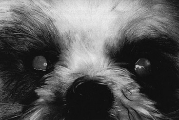
Web Chapter 77 Epiphora is defined as an overflow of tears from the conjunctival sac, as may occur with dysfunction of the lacrimal drainage system or with excessive lacrimation that results from ocular pain or irritation (Web Box 77-1). Chronic epiphora leads to staining of the facial hair and moist facial dermatitis, which results in a noncosmetic appearance and an unpleasant odor. Determination of the cause of epiphora is essential before treatment can be instituted and requires a mechanistic approach at first. Conditions that lead to excessive lacrimation should be ruled out before congenital or acquired dysfunction of the lacrimal drainage system is diagnosed. Other causes of epiphora include the irritating or painful conditions of entropion, corneal ulceration, anterior uveitis, and glaucoma (these disorders are addressed in other chapters in this section of Current Veterinary Therapy). Management of epiphora involves identifying and treating the underlying cause. When surgical treatment of the eye or adnexa is recommended to correct a cause of epiphora, referral to a veterinary ophthalmologist is recommended. Shallow orbits, as seen in brachycephalic dogs and cats, cause prominent globe positions and as a result very tight-fitting lids with small lacrimal lakes. Because of this, tears are more likely to spill over onto the face than to remain in the lake and drain through the nasolacrimal puncta and duct. These animals also have hairs that originate from the medial caruncle and canthal area and act as a wick, which facilitates drainage of tears onto the face (Web Figure 77-1). In many animals, the punctum, although normally developed and in the correct location, is occluded due to entropion of the medial lower lid or the entire medial canthal region. When the lid rolls inward, the punctum is compressed, which prevents normal tear drainage and irritates the cornea due to the resultant trichiasis. Thus careful examination of the medial canthal region and the nasolacrimal system is necessary for determination of all the factors that lead to epiphora. Web Figure 77-1 Bilateral epiphora in a 4-month-old Shih Tzu with distichiasis, trichiasis, and ectopic cilia. Chronic epiphora leads to tear staining of the facial hair. Treatment consists of surgical correction of the abnormal lid position. A Hotz-Celsus procedure can be used to correct lower medial entropion but tends to make an already large palpebral fissure even larger and may reduce the ability to blink. For this reason, a medial canthoplasty with removal of the caruncle and usually about one fourth of the upper and lower eyelid margins is performed. This corrects the macropalpebral fissure, caruncular trichiasis, and lower medial entropion while enhancing tear access to the nasolacrimal puncta (Gelatt, 2007). Cryoepilation of the caruncular hair also can be helpful if surgery is not an option. Seldom is there complete resolution of the epiphora because the shallow orbit persists.
Epiphora
Congenital Disorders
Epiphora in Small Breeds
![]()
Stay updated, free articles. Join our Telegram channel

Full access? Get Clinical Tree


