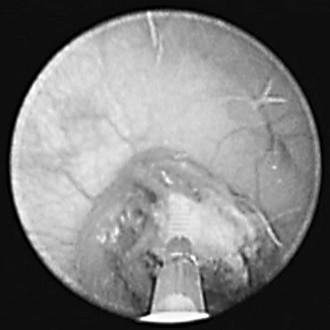
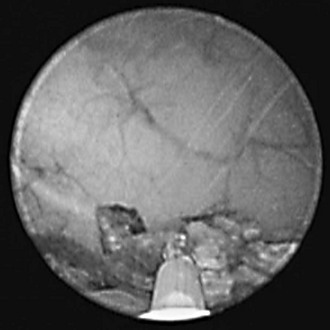
Chapter 70: Laser Lithotripsy for Uroliths

Web Chapter 70


Laser Lithotripsy for Uroliths


The term lithotripsy is derived from the Greek words lith, meaning stone, and tripsis, meaning to crush. A lithotriptor is a device for crushing or disintegrating uroliths. Extracorporeal shock wave lithotripsy commonly is used to fragment and remove uroliths from the upper urinary tract, whereas intracorporeal laser lithotripsy fragments uroliths in the lower urinary tract via urethrocystoscopy.


Successful medical application of intracorporeal lithotripsy depended on two technologic advances: (1) delivery of energy capable of fragmenting uroliths without damaging adjacent tissue and (2) development of cystoscopes capable of entering the narrow urethral lumen to visualize and manipulate uroliths and their fragments. These technologies allow fragmentation of uroliths in the urinary bladder and urethra. Urolith fragments are retrieved with a stone basket or evacuated by voiding urohydropropulsion. Urolith removal is performed cystoscopically, obviating the need for a surgical incision.


Although several forms of energy (ultrasonic, ballistic, electrohydraulic, and laser) can fragment urinary stones, not all energy forms are suitable for use in companion animals. For example, the probes for ultrasonic lithotripsy are too large to pass through the operating channel of cystoscopes commonly used for dogs and cats. The ballistic lithotripter is too rigid to traverse the curvature of the male urethra. During human treatment, the safety and efficacy of electrohydraulic lithotripsy were inferior to Holmium:YAG laser (Ho:YAG) lithotripsy. Because of the versatility and safety of laser lithotripsy and the authors’ familiarity with this treatment modality, the remaining discussion focuses on the use of Ho:YAG laser lithotripsy to manage urocystoliths and urethroliths in dogs and cats.


Mineral composition of uroliths and their size, number, and location are important considerations in the selection of minimally invasive procedures (e.g., voiding urohydropropulsion, laparoscopic-assisted cystotomy, and laser lithotripsy) for urolith management in dogs and cats (see Chapter 203 in the previous edition of Current Veterinary Therapy). Voiding urohydropropulsion may be used to remove uroliths that are smaller than the diameter of the dilated urethra (typically less than 3 to 4 mm in diameter). Laser lithotripsy effectively extends the use of voiding urohydropropulsion to larger uroliths by fragmenting uroliths into smaller pieces before transurethral evacuation (Web Figures 70-1 and 70-2). However, laser lithotripsy is not suited ideally to every patient. In small male dogs (≤5 kg) and all male cats, the lumen of the urethra may not dilate sufficiently to accommodate insertion of cystoscopies to permit urolith management.


Equipment Needed for Laser Lithotripsy


The Ho:YAG laser is the preferred laser for lithotripsy. The active medium of this laser is a crystal of yttrium, aluminum, and garnet (YAG) doped with holmium, with a wavelength of 2100 nm, which is in the near infrared portion of the electromagnetic spectrum. Because the 2100-nm wavelength of the Ho:YAG laser is absorbed in 0.5 mm of fluid, it can be used to safely fragment uroliths within the urethra or urinary bladder with minimal or no damage to the urothelium. The Ho:YAG laser energy is delivered in 350-microsecond pulses and is rapidly dispersed in water surrounding the tip of the laser fiber. Ho:YAG lasers are available with total power ranging from 20 to 100 watts, but the lower-power lasers (20 to 30 watts) are adequate for laser lithotripsy.


Laser lithotripsy can be performed through small-diameter flexible ureteroscopes (e.g., 7.5 to 10.5 Fr), which are used for urethrocystoscopy in male dogs, and rigid cystoscopes (e.g., 9 to 19 Fr), which are used in female dogs and cats. The size of the cystoscope is based on the size of the dog or cat. The laser energy of the Ho:YAG laser is transferred from the laser unit to the surface of the stone through small-diameter (200- to 550-µm) flexible quartz laser fibers. These special fibers are passed through the working channel of the cystoscope. When working through the biopsy channel of rigid cystoscope, the authors use a 365- or 550-µm quartz laser. Both 200- and 365-µm fibers pass through the 3 Fr working channel of a flexible endoscope, although care must be used to avoid damaging the working channel with the sharp rigid tip of the fiber. To protect the working channel from damage, a laser catheter can be passed over the fiber before passing the laser fiber into the scope. The smaller-diameter fibers have minimal impact on the ability to deflect the tip of flexible ureteroscopes and allow for efficient targeting of urethral calculi.


Additional equipment required for laser lithotripsy includes stone baskets used for extraction of urolith fragments. A variety of stone baskets are available from various manufacturers. The authors prefer tipless baskets that facilitate capture of small stone fragments within the urethra or bladder lumen. Although not essential, C-arm fluoroscopy is helpful to document urolith size and location during laser lithotripsy. This is most helpful in male dogs because of the limited visualization provided through flexible ureteroscopes. Fluoroscopy-aided retrograde contrast studies also can be performed to detect anatomic abnormalities associated with the uroliths and to document complete urolith removal. In female dogs and cats the entire bladder lumen can be visualized readily through the rigid cystoscope, making concurrent fluoroscopy less essential.


Laser Lithotripsy Technique


Laser lithotripsy is performed with urolith visualization by urethrocystoscopy. Detailed description of urethrocystoscopy is beyond the scope of this chapter and has been described elsewhere (Adams, 2006). The authors prefer to position female dogs and cats in dorsal recumbency and male dogs in lateral recumbency for laser lithotripsy. The area around the vulva or prepuce is clipped to remove surrounding hair and prepped for aseptic introduction of the cystoscope into the urethra and bladder. During cystoscopy irrigation of sterile warmed normal saline or sterile water is used to distend the urinary tract and facilitate cystoscope passage. Once the uroliths are visualized in the urethra or bladder, a flexible quartz laser fiber is advanced through the working channel of the scope and connected to the laser unit.


To achieve optimum fragmentation of the urolith, the quartz fiber tip must be guided with the aid of a cystoscope so that it is in direct contact with the surface of the urolith before laser activation. During laser lithotripsy continuous flushing of warmed irrigation solutions absorbs stray laser energy and flushes urolith debris and fragments out of the visual field to maintain visibility. The authors use initial power and frequency settings of 0.5 to 0.8 joules per pulse and 6 to 10 Hz for laser lithotripsy. Increasing the frequency of laser pulses (Hz) increases the efficiency of urolith fragmentation but decreases the operator’s potential reaction time in the event that the laser fiber tip inadvertently contacts the urothelium. Therefore the lowest laser settings that result in effective urolith fragmentation should be used during laser lithotripsy.


During laser activation the energy is limited to an area directly in front of the quartz laser fiber tip. An aiming beam within the visible spectrum is used to facilitate targeting the laser energy. The laser fiber should not be aimed directly at the mucosal surface and should be kept 0.5 mm or more away from the mucosal surface during laser activation. Therefore it is preferable to apply laser energy to the urolith surface while working parallel rather than perpendicular to the mucosa (see Web Figures 70-1 and 70-2). In the event of brief laser application directly to the mucosal surface, mucosal injury usually is limited to superficial damage and minimal bleeding. When in contact with the mucosal surface, the Ho:YAG laser is capable of making incisions into the bladder or urethral wall; perforation is possible during laser lithotripsy. The Ho:YAG laser can be used effectively in contact mode to remove small pedunculated polyps or tumors within the urinary tract without creating full-thickness incisions. With proper technique the risk of bladder or urethral perforation is minimal during laser lithotripsy.


Once uroliths are fractured, fragments are retrieved with stone baskets and submitted for quantitative urolith analysis. If the larger fragments were retrieved safely through the urethra, voiding urohydropropulsion is performed to evacuate remaining fragments.

< div class='tao-gold-member'>

Stay updated, free articles. Join our Telegram channel

Jul 18, 2016 | Posted by in PHARMACOLOGY, TOXICOLOGY & THERAPEUTICS | Comments Off on Chapter 70: Laser Lithotripsy for Uroliths

Full access? Get Clinical Tree

Get Clinical Tree app for offline access