Chapter 29: Nasal Tumors

Web Chapter 29


Nasal Tumors


Pathology and Clinical Presentation


Tumors of the nasal passage and paranasal sinuses in dogs account for approximately 1% to 2% of all canine neoplasms; the majority are carcinoma (60%) and sarcoma (30%) histologies. Further breakdown of histology includes adenocarcinoma, squamous cell carcinoma, fibrosarcoma, chondrosarcoma, and osteosarcoma. Dolichocephalic breeds and dogs with exposure to indoor coal or kerosene heaters are at increased risk for development of nasal tumors. Most dogs with nasal tumors are middle to older age, with reported median ages of 7.6 to 11.3 years. Nasal tumors generally are locally invasive and slow to metastasize but can metastasize to local lymph nodes (mandibular/retropharyngeal and less often to lung, abdominal organs, bone, and brain). Clinical signs in dogs with nasal tumors include nasal obstruction/respiratory stridor, sneezing, reverse sneezing, epistaxis, facial deformity, and exophthalmos. Initially signs may respond partially to antibiotics or antiinflammatory treatments because of concurrent inflammation and secondary bacterial infection that are inevitably present. These tumors often are advanced locally when diagnosed, and occasionally patients can present with neurologic signs (seizures, altered mentation, behavior change) secondary to destruction of the cribriform plate and extension into the brain.


Staging and Diagnosis


Imaging


Thoracic radiographs to evaluate for pulmonary metastases and as a general cardiac and geriatric evaluation should be obtained. Nasal radiographs can be used to image animals with nasal disease and should include the following:


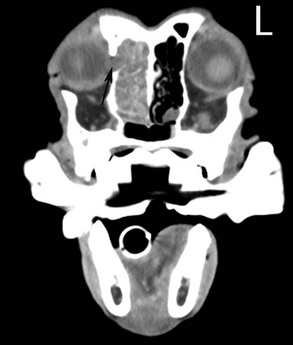
However, cross-sectional imaging is preferred. Computed tomography (CT) is the most commonly used imaging modality for evaluating dogs and cats with nasal disease, but magnetic resonance imaging (MRI) also is used. Both imaging modalities have advantages and disadvantages in the imaging of nasal disease in cats and dogs. Bone destruction is easier to identify on CT (Web Figure 29-1), and soft tissue changes are delineated better on MRI. Generally MRI studies are more expensive, and imaging time is longer than CT studies. Because computer-based radiation therapy (RT) plans are generated commonly using CT images, CT may be the preferred imaging modality if RT is contemplated.


Biopsy


Biopsy and histology of the nasal tumor are needed for definitive diagnosis. Before the biopsy procedure, additional evaluation should include complete blood count and blood chemistry along with a clotting profile. Fine-needle aspiration cytology of mandibular lymph nodes also should be performed as part of tumor staging and sometimes can yield a diagnosis in cases with lymph node metastasis. A transnostril biopsy using a closed-suction technique, a bone curette, or cup-type biopsy forceps should be performed under general anesthesia to collect a sample. Whenever a transnostril biopsy technique is used, the biopsy instrument is measured and marked to penetrate no farther than the distance from the tip of the nose to the medial canthus of the eye. This prevents penetration of the cribriform plate. A minimally invasive diagnostic and potentially therapeutic high-pressure saline hydropulsion technique also has been proposed recently. Tumor tissue generally is white; mild-to-moderate hemorrhage is expected and usually subsides within a few minutes. Cytology of nasal swabs or expectorated material is rarely rewarding. Some large nasal tumors protrude into the nasopharynx and can be sampled using retrograde rhinoscopy. When obtaining biopsy samples with smaller endoscopic instruments, clinicians also risk sampling the surrounding inflammation instead of the tumor.

< div class='tao-gold-member'>

Stay updated, free articles. Join our Telegram channel

Jul 18, 2016 | Posted by in PHARMACOLOGY, TOXICOLOGY & THERAPEUTICS | Comments Off on Chapter 29: Nasal Tumors

Full access? Get Clinical Tree

Get Clinical Tree app for offline access