Chapter 21 Cardiovascular complications in the intensive care patient
INTRODUCTION
Critically ill horses commonly develop cardiovascular complications, often associated with systemic inflammatory response syndrome (SIRS), multiple organ dysfunction syndrome (MODS) or electrolyte and acid–base disturbances that are seen in a variety of systemic conditions, most notably in the adult horse with gastrointestinal diseases and in foals with neonatal septicaemia or perinatal asphyxia syndrome. Equally, a wide range of primary cardiac disorders should be considered in horses presenting as emergencies with signs of recent onset heart failure: horses that experience sudden-onset disruption of valvular integrity can develop dramatic and fulminant signs of heart failure and horses with pericarditis or myocardial lesions such as bacterial myocarditis and cardiomyopathy also tend to present as emergencies with signs of recent onset heart failure (see Chapters 17 and 19). The clinician should also be careful not to overlook the potential for primary cardiovascular disease in animals presenting with signs of shock, pain and distress and it is important to note that horses with intrathoracic disease and certain cardiac dysrhythmias may initially display signs that are confused with colic of gastrointestinal origin. Primary cardiac diseases of the horse are extensively described in the other chapters in this book, while this chapter will focus on cardiovascular complications that are encountered commonly in the intensive care unit (ICU).
PREVALENCE AND RISK FACTORS FOR CARDIOVASCULAR COMPLICATIONS IN THE ICU
Although there are no specific data on the prevalence of cardiac problems in equine ICU, endotoxaemia is undoubtedly the major pathological state which underlies a large proportion of secondary cardiac problems seen in critically ill adult horses, while in the foal septicaemia, perinatal asphyxia and prematurity/dysmaturity are common underlying primary problems. Endotoxaemia can be associated with a range of forms of gram-negative sepsis and in adults the gastrointestinal tract is its most frequent source, while peritonitis, metritis, pleuropneumonia and neonatal septicaemia are also common potential causes. Mild fibrinous pericarditis, right atrial and ventricular enlargement, myocardial depression and ventricular tachycardia have been documented in streptococcal toxic shock with multiple organ dysfunction in a horse.1 Streptococcal toxic shock was previously associated specifically with Streptococcus pyogenes infection in humans but it is now recognized in association with a wide range of Streptococcal species in both human beings2 and dogs.3 In the last 20 years, there has been a marked increase in the frequency of diagnosis of the condition in human beings2 and it may become more important in horses in the future.1
CARDIOVASCULAR MONITORING IN THE ICU
Electrocardiography (ECG)
Clinical examination remains the most important diagnostic tool available to the equine clinician. However, in critical patients, dysrhythmias can easily go undetected on both clinical examination and using short ECG rhythm strips because dysrhythmias frequently occur intermittently and particularly in the case of monomorphic ventricular tachycardia, are often very regular and difficult to appreciate on auscultation alone. A variety of ambulatory and telemetric ECG systems are available for use in horses (see Chapter 10). Telemetric systems have the major advantage in the ICU that the clinician can identify arrhythmic episodes immediately and take appropriate action; thus, radiotelemetric ECG is the technique of choice for monitoring horses with unstable dysrhythmias and those receiving potentially pro-arrhythmic drugs.
Haemodynamic monitoring
Indirect techniques are the most accessible method for blood pressure measurement in conscious horses. The coccygeal artery is used most often in both foals and adult horses. The cuff width should be 0.2–0.26 times the tail circumference because blood pressure is underestimated if the cuff is too wide and overestimated if it is too narrow.4 The inflation bladder must be centred over the artery and the cuff fitted tightly5 to avoid inaccuracy in blood pressure measurement.6,7 The indirect blood pressure in healthy adult horses is 111.8 ± 13.3 mmHg in systole and 67.7 ± 13.8 mmHg in diastole.8 A number of studies have reported noninvasive blood pressures in healthy foals as around 80–125 mmHg in systole and 60–80 mmHg in diastole.9,10 However, the mean arterial pressure is the more important determinant of organ perfusion than the systolic or diastolic pressures and supportive intervention should be considered if the mean arterial pressures fall below 60,5,11 particularly if there are other clinical signs of poor perfusion such as abnormal mucous membranes, capillary refill time, heart rate, extremity temperature, pulse quality and urine output.12 Furthermore, while its simplicity and noninvasive nature are an undoubted advantage, the clinician should remain aware of limitations in the accuracy of indirect blood pressure measurement and therefore it is best applied as a means to identify trends within an individual patient, rather than to compare between horses.
In human ICUs, direct measurement of pulmonary artery and capillary wedge pressure is considered the most reliable means to document pulmonary hypertension and invasive measurements of cardiac output and other haemodynamic variables are widely used in monitoring and therapeutic planning. However, while technically feasible, cardiac catheterization to allow direct measurement of pulmonary artery pressure or facilitate cardiac output measurement is still rarely employed in equine patients. ( PH) Nevertheless, practical means to measure cardiac output and other haemodynamic indices, such as lithium dilution, are becoming more widely available and are likely to be used in equine critical patients with increasing frequency.13,14 Monitoring of the cardiac output, stroke volume and systemic vascular resistance can allow the clinician to distinguish whether the cardiovascular status is compromised by reduced circulating volume, vascular tone, cardiac output or a combination of these and in doing so, select the most appropriate treatment and monitor the effects of vasopressors, inotropes and volume expansion.5,11
Echocardiography
Echocardiography is the most valuable diagnostic aid for assessment of cardiac structure in horses and thus is the technique of choice in horses presenting with cardiac murmurs, and it is useful in ruling in or out structural heart disease in horses presenting with cardiac dysrhythmias. In the ICU setting, although echocardiography has the potential to assess cardiac output and pulmonary hypertension (see Chapter 9), it is less accurate than the gold-standard direct methods. On the other hand, its noninvasive nature is particularly attractive. Due to limitations in their accuracy, echocardiographic measurements of haemodynamic indices should be used primarily to identify trends within patients rather than taken as absolute measurements. Furthermore, the distributive shock that occurs with endotoxaemia and SIRS is principally due to dysregulation of systemic vascular function and accompanied by microthrombosis and possibly a direct myocardial depressant mechanism. Echocardiography is not a particularly useful tool for identifying these haemodynamic alterations when compared to measurement of cardiac output by thermodilution or lithium dilution, although horses and foals with endotoxaemia or septicaemia often have nonspecific echocardiographic signs of global cardiac dysfunction such as reduced fractional shortening, spontaneous contrast and poor ventricular wall movement. Hypovolaemic patients can have reduced cardiac chamber size and in septicaemic patients mild pericardial effusions are fairly common.1
SPECIFIC FORMS OF CARDIAC COMPLICATIONS IN INTENSIVE CARE PATIENTS
Circulatory collapse
Circulatory collapse, or shock, can arise from reduced cardiac output, reduction of circulating volume, dysregulation of vasomotor tone or a combination of these factors. Acute heart failure is described in Chapter 19. Endotoxaemia, sepsis and SIRS are classified as forms of distributive shock as they are characterized by widespread systemic vasodilation although both hypovolaemia and myocardial depression can contribute to the pathogenesis. Hypovolaemia can be due to severe haemorrhage or sequestration of fluid into the body cavities or into the gastrointestinal or urogenital tracts. The main goals of therapy in circulatory collapse are to restore and maintain the circulating volume and to attempt to increase vasomotor tone and cardiac output with pressor and ionotropic agents (see Chapter 7). Clearly, treatment aimed at addressing and correcting the predisposing condition is critical. Global myocardial depression is difficult to document in equine critical care patients but the techniques described above can be very helpful in identifying cardiac abnormalities that accompany circulatory collapse.
Cardiac dysrhythmias
In horses, dysrhythmias occurring secondary to other systemic diseases, particularly gastrointestinal disease,15,16 are encountered more frequently than rhythm disturbances associated with primary myocardial pathology. In a group of 67 horses with duodenitis/proximal jejunitis, six horses (9%) had dysrhythmias detected by auscultation and conventional electrocardiography.15 The prevalence of dysrhythmias in the postoperative period following abdominal surgery is not known but is likely to be higher than may be appreciated on auscultation alone: in one survey of horses with ventricular dysrhythmias, 4 of 21 cases had recently undergone colic surgery16 and ambulatory ECGs obtained from fifty horses within 3 days of exploratory celiotomy demonstrated that 11 horses (22%) had isolated supraventricular premature depolarizations while 8 horses (16%) had ventricular dysrhythmias, including 4 (8%) with idioventricular rhythms or paroxysmal monomorphic ventricular tachycardia.17 In all but one of these cases the dysrhythmias were self-limiting, no specific treatment was required and the dysrhythmia had not been recognized on auscultation. Occasionally, more clinically significant dysrhythmias are encountered in the postoperative period following colic surgery, and in this instance clinical signs of reduced cardiac output and marked tachycardias may be apparent. A high index of suspicion is required and the clinician should consider an ECG assessment in all horses that have heart rates that are higher than might otherwise be suspected given the degree of pain or in horses showing weakness, pallor and other signs of low cardiac output.
Causes of dysrhythmias secondary to gastrointestinal disease include the direct effects of endotoxin on the myocardium, autonomic imbalance resulting from gastrointestinal distension and metabolic, electrolytic or acid–base imbalances.15 Ideally, underlying and contributory factors should be addressed first. The decision to institute specific antidysrhythmic therapy is generally based on assessment of whether it is likely that the dysrhythmia will destabilize into a life-threatening state. In general, ventricular dysrhythmias are much more likely to require antidysrhythmic therapy than supraventricular dysrhythmias, and commonly applied guidelines suggest that antidysrhythmics should be considered where the heart rate is rapid (greater than 100 bpm), the dysrhythmia is polymorphic and R on T phenomenon is present (see Fig. 13.17). However, the most important thing to consider is the clinical status of the animal, and the decision of whether to utilize antidysrhythmics should be based on the presence or absence of signs of low cardiac output. Strong evidence to support decisions on which specific antidysrhythmic agents to use in equine patients is lacking but procainamide, lignocaine and quinidine gluconate are popular first choices in ventricular tachycardia. Magnesium sulphate has been successful, and can be used alone, or in combination with other antidysrhythmic agents. Further details of these and other antidysrhythmics are described in more detail in Chapter 13. ( VT)
VT)
The electrolytes that are most commonly associated with dysrhythmias are potassium, calcium and magnesium and the role of these electrolytes in arrhythmiogenesis is discussed in detail in Chapter 6. Hypokalaemia, hypocalcaemia and hypomagnesaemia are common in horses with sepsis and endotoxaemia,18 thus monitoring and addressing electrolyte imbalances in critically ill horses is an important priority.
Hypokalaemia
Hypokalaemia is commonly found in ill animals and causes include endotoxaemia,18 anorexia, diarrhoea and starvation.19 It is frequently present in horses with heat exhaustion along with hypochloraemia, hypocalcaemia and metabolic alkalosis. Hypokalaemia leads to prolongation of the Q–T interval and both supraventricular and ventricular dysrhythmias can be seen in horses with hypokalaemia. Supraventricular tachycardia, ventricular tachycardia, torsades de pointes and ventricular fibrillation can all occur with severe hypokalaemia. If severe hypokalaemia is present, the calculated potassium deficit should be replaced slowly intravenously at a maximum rate of 0.5 mEq/kg/hour while monitoring serum potassium concentrations.
Hyperkalaemia
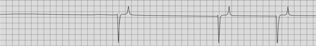
Hyperkalaemia is uncommon in horses except in foals with uroperitoneum. Hyperkalaemia is occasionally detected in adult horses, primarily those with acute renal failure or in Quarter horses with hyperkalaemic periodic paralysis (HYPP); this condition is described in more detail in Chapter 20. Cardiac dysrhythmias may or may not be detected on auscultation in hyperkalaemia but an ECG should be obtained in horses or foals with a plasma potassium equal to or greater than 6 mEq/L. Tall peaked T waves are detected with plasma potassium values equal to or greater than 6.2 mEq/L (Fig. 21.1). Progressive slowing of conduction and decreased excitability occur with hyperkalaemia and may result in cardiac arrest or ventricular fibrillation. Broadening and flattening of the P waves, prolonged P–R intervals and bradycardia are a reflection of this on the ECG. Atrial arrest or atrial standstill develops with progressive hyperkalaemia. Supraventricular and ventricular premature depolarizations and ventricular tachycardia have also been reported. Widened QRS complexes are further indications of severe, near lethal hyperkalaemia.
< div class='tao-gold-member'>
Stay updated, free articles. Join our Telegram channel

Full access? Get Clinical Tree


