Chapter 3 Cardiac responses to exercise and training
INTRODUCTION
Heart rate and cardiac output increase quickly at the commencement of exercise, enabling comparatively rapid increases in the rate of oxygen consumption. The 6- to 8-fold increase in heart rate during maximal exercise, coupled with an increase in stroke volume, results in maximal cardiac output during exercise of approximately 300 L/minute, or over 0.6 L/minute/kg in trained horses. This large increase in blood flow to exercising muscle is fundamental to the athletic performance of the horse during intense exercise. This is especially so in events of more than 1 minute duration, which rely mainly on aerobic energy supply.1 Training results in adaptations that increase the rate of blood flow to skeletal muscle during intense exercise. The main adaptation is an increase in stroke volume. Training also causes changes in the electrocardiogram and echocardiogram.
CARDIAC RESPONSES TO EXERCISE
Heart rate
Heart rate during exercise
Heart rate quickly increases at the commencement of exercise, and reaches a steady state in 2–3 minutes. This increase is associated with increased sympathetic nerve activity and/or catecholamine release.2 An overshoot of heart rate to levels above the submaximal steady-state heart rate may occur at the commencement of exercise. Thereafter the heart rate gradually decreases to a steady state.3
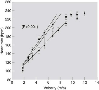
A linear relationship between heart rate and submaximal work effort has been observed in horses trotting, galloping and swimming.3–8 This relationship is usually defined by use of treadmill or racetrack exercise tests which involve increasing the speed every 1–2 minutes and measuring the heart rate at the completion of exercise at each speed. After a suitable warm up, for example, 3 minutes trotting, the heart rate is stable after 1 minute of further exercise at higher speeds. Heart rates recorded during an exercise test in three groups of Thoroughbred horses are illustrated in Figure 3.1.8 The figure demonstrates the linear relationship between heart rate and speed of submaximal exercise. At higher speeds, further increases in treadmill speed are not accompanied by an increase in heart rate, and a plateau occurs. The horse is then exercising at its individual maximal heart rate.
 ), two-year-old (
 ) and adult (•) Thoroughbred horses exercising on a treadmill set at 6 degrees; Yearlings: n = 16, y = 93.30 + 19.44x, r2 = 88%. Two-year-olds: n = 12; y = 89.85 + 18.05x, r2 = 80%. Adults: n = 14; y = 79.99 + 14.60x, r2 = 92%.
The typical relationship between heart rate and speed of exercise enables variables such as V200 and VHRmax to be calculated. These measurements have been used to compare different groups of horses, or individual horses with a normal group, or measure changes in the heart rate response to exercise during training or detraining.9 V200 is the velocity which generates a heart rate of 200 beats per minute. VHRmax is the velocity at which the loss of linearity occurs and no further increase in heart rate occurs, despite an increase in velocity. Derivation of these variables is illustrated in Figure 3.2.9 Some studies have also used V140, the velocity that results in a heart rate of 140 beats per minute. It has been argued that V200 is superior because it approximates the intensity of exercise which results in a blood lactate concentration of 4 mmol/L.10 However, this argument should not dictate strict adherence to V200. If V200 is used to compare the fitness of different horses, the measurement could be very misleading. Two horses could have the same V200, but very different maximal heart rates, and therefore very different velocities at which they reach their maximal heart rates. The V200 ignores the cardiac reserve between 200 bpm and maximal HR, and so is best used to measure changes in an individual horse over time, assuming that maximal HR does not change.
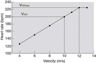
Figure 3.2 Determination of V200 and VHRmax from measurements of heart rate at treadmill speeds of 4, 6, 8, 10, 11, 12 and 13 m/s (treadmill slope 10%).9
The heart rate versus work relationship during treadmill exercise is very reproducible for individual horses at heart rates between 120 and 210 bpm.5,11 During racetrack exercise, the reproducibility of V200 in Standardbred horses measured in a racetrack exercise test was also good.12 This study also found that V200 was not significantly altered by a change of racetrack on which the test was conducted. However, in an individual horse extraneous factors can influence the heart rates during an exercise test. For example, anxiety, the presence of a breathing mask10 and changes in treadmill exercise testing routine13 can elevate heart rate during exercise.
The normal heart rate responses to treadmill exercise in Thoroughbred horses have been described.8,14 Figure 3.1 illustrates the responses in 16 yearling, 12 two-year-old and 14 trained adult Thoroughbreds.8 The slope of the linear regression of heart rate on m/s in the trained adult horses was significantly less than the slopes for both the untrained two-year-olds and yearlings.
There is a linear relationship between heart rates and oxygen consumption during submaximal exercise (Fig. 3.3A15). This relationship is not affected by the slope of the treadmill in the range 0–10%. Likewise, there is a linear relationship between relative heart rate and relative oxygen uptake during submaximal exercise (expressed as percentages of maximal values) (Fig. 3.3B15). This close relationship infers that the velocity at which an individual horse attains its maximal oxygen uptake is equivalent to the VHRmax..
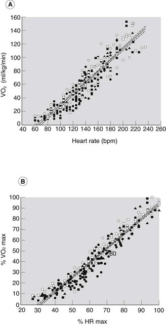
Figure 3.3 Relationships between oxygen uptake ( ) and heart rate (HR) (A) and between % HRmax and %
  (B) in five Thoroughbred racehorses during submaximal treadmill exercise. Results are pooled from exercise tests conducted at 0, 2.5, 5, 7.5 and 10% treadmill inclines.15 •, 0%;
  , 2.5%;
  , 5%;
, 5%;  , 7.5%;
  , 10%. A:
  = 0.833 (HR) – 54.7, r2 = 0.865. B: %
  = 1.348 (% HRmax) – 41.0, r2 = 0.939.15
The overshoot at the commencement of exercise may be absent if the exercise is at a high intensity. Instead, heart rates may gradually increase to maximal. Overshoot of heart rate occurred at the start of exercise at 50% of  (maximum oxygen uptake) in Standardbred horses. However, at 100%
  , heart rate gradually increased during a 5 minute period of exercise.16 The slow increase in heart rate towards the true maximal heart rate in that study may have been related to the absence of a warm up period of exercise.
Heart rates during swimming vary greatly between horses, and are usually in the range 130–180 bpm.6,17 The mean highest heart rate recorded in nine horses during show jumping were 191 ± 3 bpm.18 Mean (±s.d.) heart rates during phases A and C of an advanced 3-day event were 118 ± 11 and 135 ± 28, respectively. During phases B and D, heart rates were 175 ± 23 and 171 ± 19, respectively. A continuous record of heart rates in a horse competing in the event is illustrated in Figure 3.4.19
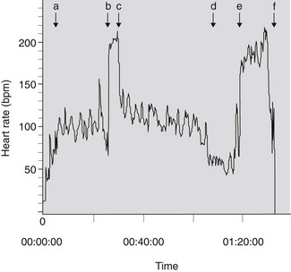
Figure 3.4 Heart rate of a horse competing in an advanced (CCI***) three-day event. aDenotes the beginning of phase A, bdenotes the beginning of phase B, cdenotes the beginning of phase C, ddenotes the end of phase C and beginning of 10-minute box, edenotes the beginning of phase D and fthe end of phase D.19
During prolonged strenuous submaximal exercise at a constant work rate, a gradual increase in heart rate, or “cardiovascular drift” can occur. For example, during 30 minutes of exercise, mean heart rate increased from 154 to 173 bpm.20 This “drift” was accompanied by increases in minute ventilation and stroke volume, while cardiac output was unchanged. In another study, horses exercising at 55–60% of individual HRmax for 60 minutes had minimal changes in heart rate.21 Heart rate responses during prolonged exercise probably depend on hydration status and the environmental conditions.
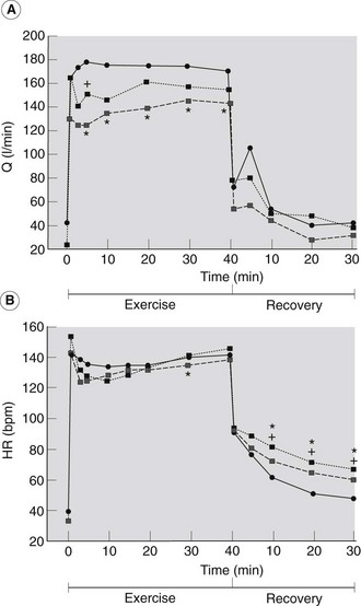
The effects of dehydration on heart rate during and after 40 minutes of exercise eliciting approximately 40% of maximal oxygen consumption have been described.22 Horses were exercised in three conditions; euhydrated, 4 hours after administration of furosemide (1.0 mg/kg i.v.) to induce isotonic dehydration, and after 30 hours without water to induce hypertonic dehydration. The most pronounced heart rate drift occurred in the dehydrated horses. Heart rates after 30 minutes of exercise were significantly higher in the horses dehydrated by treatment with furosemide than in the control, euhydrated condition. During the recovery period heart rates were also higher in both groups of dehydrated horses than in the control horses (Fig. 3.5). This study demonstrated that dehydration results in higher heart rates during and after prolonged submaximal exercise.
 ——
—— ) and by water deprivation (DDH
  ……..
…….. ). *FDH significantly different from C; +DDH significantly different from C22.
Diseases such as chronic obstructive pulmonary disease result in heart rates during submaximal exercise that are significantly higher than those found in normal horses.23–25 Measurements of heart rate and other cardiorespiratory and metabolic indices during treadmill exercise have also been used to investigate the functional significance of various electrocardiographic findings. This technique was used to illustrate that T wave changes and second degree AV block in the resting electrocardiogram do not result in significantly different heart rates or peak oxygen consumption in Thoroughbred and Standardbred racehorses.26
High heart rates during submaximal exercise are also to be expected in horses with diseases which limit stroke volume, such as atrial fibrillation.27 ( AF) When valvular dysfunction, or structural cardiac disease results in cardiac decompensation, affected horses will also have an increased heart rate during exercise, before clinical signs of heart failure are evident at rest.28
However, lameness should also be considered as a cause of high heart rates during submaximal exercise tests. Mild unilateral forelimb lameness also causes lower maximal oxygen uptake and more rapid blood lactate accumulation during intense exercise.29 High heart rates could also be due to anxiety if the horse has not been adequately acclimated to the treadmill test procedures. Field studies of submaximal heart rate could be easier to interpret because the horse is exercising in its usual environment, and should be less subject to the effects of fear or anxiety.
The highest heart rates recorded during racing in 19 Thoroughbreds averaged 223 bpm, with a range of 204–241 beats/minute.30 Mean peak heart rate in eight yearling Thoroughbreds was approximately 240 beats/minute, compared to 220–230 in 2- to 4-year old horses.14 Likewise, yearling, two-year-old and adult Thoroughbreds had similar means (229–231 bpm) and ranges (215–254 beats/minute) of peak heart rates during an incremental treadmill exercise test.8 In a recent retrospective multicentre study, horse age, gender, breed, use and fitness combined with the testing centre itself were shown to affect peak heart rate in 394 horses undergoing standardized treadmill exercise tests.31 High interindividual variability in heart rates while racing have also been recorded in Standardbreds, ranging from 210 to 238 with a mean of 221 beats/minute.32 However, in an individual horse, the maximal heart rate is highly repeatable.11 Further work is required to confirm that the highest (peak) HRs recorded during Thoroughbred and Standardbred races are equal to the maximal HR attainable by the horse.
A new technique has been established for the study of submaximal and maximal heart rates during field gallops in Thoroughbred racehorses. This approach uses simultaneous measurement of velocity with a global positioning system, and heart rates. Derived indices of fitness such as V200 and VHRmax during the field gallop on a racetrack have been measured reliably, and have demonstrated increases with training in 2-year-old Thoroughbreds33 VHRmax has also been correlated with retrospective racing performance in Thoroughbreds.34
Heart rate after exercise( AF)
AF)
Heart rate recovery is usually very rapid in the first minute after exercise stops.3,35–37 It then decreases more gradually towards normal resting values. The rate of heart rate recovery after maximal treadmill exercise is probably independent of training state,8 although clinical experience suggests that the rate of heart rate recovery does seem to be related to conditioning in a broader range of equine athletes under field conditions. In common with heart rates at rest and during submaximal exercise, heart rates during recovery are also susceptible to rapid fluctuations due to excitement.35 Heart rates during recovery from the cross country (Phase D) of a 3-day event were not correlated with heart rates during competition.38
After prolonged submaximal exercise, recovery heart rates are higher in horses that were dehydrated before exercise.22 Poorly performing endurance horses have higher postexercise heart rates than the better performers.39 Horses with heart rates less than 60 beats/minute at 30 minutes post-exercise had less evidence of dehydration and myopathy.40 Horses with heart rates greater than 65–70 beats/minute at the 30 minute recovery time at the mid-point of an endurance ride were more prone to develop severe dehydration and exhaustion when allowed to continue.41
Most studies report that heart rate in resting horses does not decrease after training,42–45 but decreases do occur during submaximal exercise.46,47 However, the decrease in heart rate with training is often only 10–20 beats/minute at any submaximal speed.42 Some studies have also reported that heart rates during submaximal exercise were not significantly different after training.44,47 In two treadmill training studies which demonstrated significant increases in maximal oxygen consumption, no significant changes were found in heart rate during submaximal exercise.44,48 Decreasing heart rate during submaximal exercise is therefore an unreliable index of fitness in horses. Perhaps horse excitement, or anxiety, during treadmill exercise tests, renders measurements such as V200 less reliable, compared to similar measurements made in the field in a more familiar environment.33
The individual HRmax is not affected by training, and it is not a useful measure of fitness.8,47,49 Individual HRmax can be affected by the cardiac rhythm abnormality, atrial fibrillation.28 During atrial fibrillation peak heart rates during exercise are greatly increased compared to during normal sinus rhythm. In clinical cases affected by paroxysmal or sustained atrial fibrillation, heart rates during submaximal exercise can approach 300 beats/minute. As a result of the abnormally high heart rates maximal stroke volume is decreased due to suboptimal ventricular filling, and peak athletic performance is adversely affected. Resolution of the abnormal rhythm after treatment or spontaneous conversion usually results in return to normal individual HRmax and previous levels of athleticism.
Postexercise heart rates have shown no significant changes due to training in several studies.44,45,48 However, heart rates taken within 1–5 minutes of completion of fast work by Thoroughbreds were lower after training in another study.47 Analysis of fitness using postexercise heart rates may be limited because of the rapid cardiac deceleration after cessation of exercise and the influence of psychogenic factors at heart rates less than 120 beats/minute.10
Echocardiography has been used to study cardiac function after treadmill exercise.50 The heart rate recovery appeared to be prolonged after exercise in hot humid conditions (30°C and 80% relative humidity) compared to a recovery after exercise in a cooler environment. Echocardiographic indices of ventricular dimensions and contractile performance were also lower after exercise in hot humid conditions. It was suggested that these responses could reflect a reduction in preload due to the effect of dehydration or the redistribution of blood flow. If echocardiography is used to clinically evaluate cardiac function after exercise as has been suggested51 the exercise test and environmental conditions must both be strictly standardized.
Stroke volume and cardiac output
Stroke volume in the resting horse is approximately 800–900 mL, or about 2–2.5 mL/kg.42,43 Stroke volume increases by about 20–50% in the transition from rest to submaximal exercise.7,46,52,53 It does not change as intensity of exercise increases from approximately 40%  to 100%
  , despite the limited time available for ventricular filling at high heart rates during exercise.54 Stroke volumes of 2.4 mL/kg (1250 mL)54 and 3.8 ± 0.4 mL/kg (approximately 1700 mL)55 have been reported in fit Thoroughbreds during treadmill exercise at
  . This large difference could reflect biological variation, or differences in the method of measuring oxygen uptake during the exercise test.
Values reported for cardiac output in fit Thoroughbreds during treadmill exercise at  are 534 ± 54 mL/kg/minute (277 L/minute)54 and 789 ± 102 mL/kg/minute (355 L/minute).55
During tethered swimming at low work loads, stroke volume decreased from 2.06 mL/kg at rest to about 1.5 mL/kg. This response may be related to decreased venous return secondary to the alterations to breathing pattern during swimming.6
During prolonged exercise cardiac output is decreased in dehydrated horses, and this limits thermoregulation.22 In an experiment in which horses were exercised for 40 minutes while euhydrated, or dehydrated by either withdrawal of water (DDH) or administration of furosemide (FDH), cardiac output was significantly lower in FDH (144.1 ± 8.0 L/minute) and in DDH (156.6 ± 6.9 L/minute) than in euhydrated horses (173.1 ± 6.2 L/minute) after 30 minutes of exercise (see Fig. 3.5). Dehydration resulted in higher temperatures in the middle gluteal muscle and pulmonary artery during exercise, but temperatures in the superficial thoracic vein and at subcutaneous sites on the neck and back were not significantly different. Sweating rates were also similar in control and dehydrated horses, and it was concluded that the impairment of thermoregulation was primarily due to decreased transfer of heat from core to periphery.
Stay updated, free articles. Join our Telegram channel

Full access? Get Clinical Tree


