Ataxia

CHAPTER 132 Ataxia


Ataxia, or incoordination of gait, is frequently associated with concurrent clinical signs of spasticity (stiffness) and paresis (weakness) and may be a result of diffuse disease or a focal or multifocal lesion anywhere in the central nervous system (CNS), including the cerebrum, cerebellum, brainstem, or spinal cord (Table 132-1). Cerebral, cerebellar, and brainstem lesions are uncommon but can result in ataxia in addition to other clinical signs referable to lesions in those areas. In this chapter, common conditions associated with ataxia are reviewed.
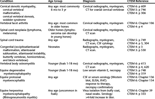


INTRACRANIAL CONDITIONS


With a unilateral cerebral lesion, contralateral proprioceptive deficits of the thoracic and pelvic limbs, hemiparesthesia, and contralateral cortical blindness may be observed. Depending on the size of the cortical lesion, mentation may be normal, but as the size of a lesion increases, there will be increasing depression and a decreased level and quality of consciousness. Dementia and seizures confirm the presence of a cortical lesion. The horse may circle compulsively, with the neck bent toward the side of the lesion.


Cerebellar disturbances result in ataxia and an inability to regulate the rate, range, and force of movement. Horses often have dysmetria; either hypermetria, with higher and longer flight of the limb, or hypometria. Initiation of movement may be jerky, and truncal sway is common. Spasticity caused by hypertonia results in stiff, jerky movements. Cerebellar lesions do not cause paresis, although hypotonia can make a horse appear weak or drag its toes. Intention tremor is obvious with severe diffuse cerebellar disease. Unilateral lesions result in signs ipsilateral to the lesion.


Ataxia associated with vestibular disease is usually asymmetric, and usually strength is preserved. Ataxia results when the head does not orient with movement of the eyes, trunk, and limbs, resulting in loss of balance. Horses with vestibular ataxia lean, drift, or fall toward the side of the lesion and usually have ipsilateral head tilt combined with nystagmus. Vestibular lesions may be central or peripheral. Horses with signs of both vestibular ataxia and facial paralysis are most likely to have otitis or stylohyoid osteoarthropathy.


CONDITIONS INVOLVING THE SPINAL CORD


In the absence of brain or brainstem disease, ataxia involving all four limbs is most commonly caused by a diffuse or multifocal cervical spinal cord lesion. Clinical signs are commonly more obvious in the pelvic than in the thoracic limbs. Head elevation can worsen spasticity, resulting in floating of the thoracic limbs and pacing, which is a significant finding in nongaited breeds. Turning the horse in tight circles exacerbates signs of proprioceptive deficits, and wide spastic circumduction of the outer limb, pivoting on the inside limb, and toe-dragging and interference, with the horse stepping on itself or striking the medial aspect of the limbs, are common. Subtle abnormalities can be worsened by repeating the procedure with the head slightly elevated or with the horse circling while on a slight slope.


Horses with cervical lesions may have difficulty backing up, with the limbs protracted slowly or dragged back in an awkward fashion. Weakness can be evaluated subjectively by assessing the horse’s resistive strength and recovery of balance when the tail is pulled sharply to one side during walking. The tail should be pulled to both sides, and the timing of the tail pull should coincide with weight bearing on the ipsilateral hind limb. Sometimes the horse will stumble in attempts to correct its posture. Horses with a lesion high in the cervical portion of the spinal cord (C1-2) may also have a head tilt and may lean to one side, mimicking vestibular disease, although no pathologic nystagmus is present and no loss of balance is observed when the horse is blindfolded. A lesion in the thoracic intumescence (C6-T2) can cause ataxia of all four limbs, accompanied by lower motor signs of denervation atrophy, weakness, and possible paraesthesia of certain sensory cutaneous areas of the thoracic limbs. Clinical signs restricted to the pelvic limbs result from thoracolumbar lesions.


CERVICAL VERTEBRAL STENOTIC MYELOPATHY


Cervical vertebral stenotic myelopathy (CVSM) is also known as cervical vertebral malformation, cervical vertebral stenosis, cervical vertebral instability, and so-called wobbler syndrome. CVSM can develop in any horse at any age but is typically seen in young, fast-growing horses. The condition is most commonly reported in Thoroughbreds (up to 2%) and Quarter Horses and is more frequent in males than in females.


Bone malformation of the cervical vertebrae leads to narrowing of the cervical vertebral canal, which results in spinal cord compression and development of neurologic signs. CVSM can be divided into two forms: static and dynamic spinal cord compression. In horses with dynamic stenosis, compression of the spinal cord during a myelogram is observed only during movement of the neck, particularly during flexion (see Chapter 131, Diagnostic Imaging in Neurologic Disease). Dynamic compression primarily affects horses from 6 to 18 months of age and most commonly occurs at C3-4, followed in frequency by C4-5. During myelography, static cervical stenosis can be observed as a narrowing of the vertebral canal and resultant spinal cord compression at any neck position, although the compression is often exacerbated in the extended views of the neck. Static stenosis is most commonly observed at the C5-6 and C6-7 articulations in horses aged 1 to 4 years, but it is also seen in older horses. Horses older than 4 years often have clinically significant vertebral facet arthritis that can lead to further compression of the spinal cord, signs of neck pain, and even impingement on the spinal nerves as they exit the intervertebral foramina. This can lead to signs of lower motor neuron disease, including atrophy of cervical musculature, cutaneous hypalgesia, or focal sweating.


Pathogenesis


The condition has a multifactorial etiology and is associated with overfeeding and other nutritional imbalances, especially of calcium, phosphorus, copper, and zinc. Cervical vertebral stenotic myelopathy may be a form of osteochondrosis involving the epiphyseal plate or articular processes. Vertebral instability leads to muscle and ligament hypertrophy, which can also lead to spinal cord compression. Prolonged compression or repetitive traumatic injury to the spinal cord initially results in damage to the peripheral white matter. The proprioceptive and spinocerebellar tracts of the pelvic limbs are positioned more superficially in the spinal cord than those of the thoracic limbs, which might explain why more severe ataxia is observed in the pelvic limbs compared with the thoracic limbs.


A hereditary predisposition is likely, but this point remains controversial. In a study in which wobbler stallions were bred to wobbler mares, there was a high incidence (45%) of foals with osteochondrosis but no increased incidence of wobblers. Nonetheless, certain stallions are known to sire a high proportion of wobbler foals. Breeding of wobbler stallions and mares is therefore best avoided.


Young horses often manifest acute onset of ataxia or gait abnormalities, often associated with a fall. In many instances, the horse was mildly ataxic before the traumatic episode, and the mild neurologic deficits may even have been a predisposing cause of the fall. The traumatic incident exacerbates the neurologic signs because of cord swelling within the stenotic vertebral column.


May 28, 2016 | Posted by in EQUINE MEDICINE | Comments Off on Ataxia

Full access? Get Clinical Tree

Get Clinical Tree app for offline access