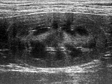
Applications of Ultrasound in Diagnosis and Management of Urinary Disease

Chapter 185


Applications of Ultrasound in Diagnosis and Management of Urinary Disease


Diagnostic imaging traditionally has played an important role in the workup of veterinary patients with disorders of the urinary system. With increases in technical quality and operator experience, ultrasonographic examinations and ultrasound-guided procedures have become invaluable tools and have partially replaced more conventional techniques such as survey radiography and radiographic contrast procedures. Ultrasound is useful for the diagnosis of various disorders of the kidneys, ureters, urinary bladder, and urethra; allows controlled needle placement for further diagnostic and therapeutic procedures; and helps in monitoring a patient’s response to treatment.


Indications and Limitations


Ultrasound not only is indicated in patients with signs related to the urinary tract such as hematuria, stranguria, and increased frequency of urination but also is useful in the assessment of patients with nonspecific clinical signs such as abdominal pain or palpable abdominal masses. It is excellent for the evaluation of diffuse or focal renal parenchymal lesions, renal pelvis and ureteral dilation, intraluminal and mural lesions of the urinary bladder, and abnormalities of the intraabdominal part of the urethra. However, when one is deciding which imaging modality to choose for a patient with suspected urinary tract disease, the limitations of ultrasound need to be taken into consideration. In general, the diagnostic quality of abdominal ultrasound may be limited in large, deep-chested, and obese patients and may be affected significantly by the presence of a large amount of gas or ingesta within the gastrointestinal tract. Normal (nondistended) ureters and the intrapelvic part of the urethra typically are not visible when an ultrasonographic examination is performed and, depending on the indication, usually are better examined by means of an excretory urogram or a urethrogram, respectively. Ultrasound is inferior to survey radiography in the diagnosis of mineral opaque calculi in ureters and urethra, in the determination of the exact number of cystic calculi, and in the verification of the number of kidneys in the rare case of missing or supernumerary kidneys. Finally, contrast procedures such as excretory urography and cystourethrography are superior in the diagnosis of ureteral or urinary bladder rupture, respectively; in the identification of the bladder in cases in which numerous large fluid-filled mass lesions are present in the caudal abdomen; and in the assessment of most urethral lesions.


Technique


The choice of transducer type and frequency is dictated in most instances by the equipment available in the examiner’s practice. A microconvex, multifrequency transducer is desirable because it has a small contact area with the skin, allows the right kidney to be imaged through an intercostal approach without interference from overlying ribs, and provides the option of adjusting the transducer frequency during the examination. Curvilinear or linear transducers may be used if a microconvex transducer is not available. The transducer frequency should be chosen based on patient size and depth of the organs to be imaged. In large-breed dogs a frequency as low as 5 to 8 MHz may be needed to image the kidneys, whereas kidneys in small animals and the urinary bladder can be imaged with a higher frequency (8 to 12 MHz). As in the examination of other organ systems, the highest transducer frequency allowing evaluation of an organ should be chosen for each patient, and frequency may need to be adjusted separately for kidneys and urinary bladder.


The technique for the ultrasonographic evaluation of the urinary tract follows the same principles as that for a general abdominal examination. The animal is positioned in dorsal (or, depending on examiner preference, lateral) position, the abdomen is clipped, and ultrasonographic coupling gel is applied. The left kidney and urinary bladder usually are visualized easily using a ventral abdominal approach, as are dilated ureters and the proximal part of the urethra. The right kidney may be difficult to image because of its position in the craniodorsal abdomen immediately caudal to the liver, especially in large-breed and deep-chested dogs. In these cases use of a right dorsal intercostal approach, with the patient in left lateral recumbency, is beneficial to visualize the kidney and proximal right ureter. In male dogs the penile urethra can be scanned via a perineal and ventral approach.


The majority of the ultrasonographic examination is performed using routine grayscale B-mode ultrasound. Color Doppler or power Doppler examination is helpful in assessing blood flow to an organ (e.g., in cases of suspected renal infarction) and in distinguishing vascular lesions such as bladder or renal neoplasms from nonvascularized lesions such as blood clots in the urinary bladder or renal cysts. Spectral Doppler ultrasonography and measurement of specific hemodynamic parameters such as resistive and pulsatility indices are not performed commonly in veterinary patients because of their low diagnostic yield in common urinary tract disorders.


If an ultrasound-guided interventional procedure is to be performed after the scan, all ultrasound gel should be cleaned off the abdomen. For simple procedures such as cystocentesis, thorough cleaning of the abdominal wall with alcohol is sufficient. When more invasive procedures such as renal biopsy are performed, surgical preparation of the abdominal wall is indicated. Cystocentesis and fine-needle aspiration of a kidney in a small patient can be accomplished using a 1.5-inch 22-gauge needle and syringe. However, aspiration of deeper structures necessitates the use of longer needles (e.g., 2.5-inch or 3.5-inch spinal needles). When long needles are used needle placement is more difficult to monitor with ultrasound. For these cases, and for biopsies utilizing larger bore sizes or extended procedures (such as drainage of large cysts), use of needle guides is strongly recommended. The guides consist of probe attachments and needle channels that direct the intraabdominal course of the needle in a predetermined direction and thus minimize the risk of injury to surrounding structures.


Normal Findings


Normal kidneys have an ovoid shape and are smoothly marginated. Generally, the size of the left and right kidney in the same animal should be similar. Because of the large variability in size between dogs of different breeds renal size traditionally has been evaluated with radiography rather than ultrasound. A normal range of 5.5 to 9.1 for kidney : aorta ratio has been proposed recently that allows objective assessment of renal size in a given dog. In cats, renal size is more consistent and should be approximately 3.0 to 4.5 cm in length. The renal cortex is hyperechoic to the strongly hypoechoic centrally located medulla (Figure 185-1), hypoechoic to the spleen, and isoechoic to hypoechoic to hepatic parenchyma. Many factors affect the echogenicity of the renal cortex, including normal variation, renal fat deposition, and transducer frequency so care must be taken not to overinterpret a mild generalized increase in renal cortical echogenicity as a pathologic change. The medulla is hypoechoic, and interlobar and arcuate vessels can be followed as anechoic tubular structures with hyperechoic walls crossing the renal medulla and branching at the level of the corticomedullary junction, respectively. When the kidney is investigated with color Doppler ultrasonography additional smaller branches (interlobular vessels) may be seen in the renal cortex. The renal pelvis may or may not be visible and should measure less than 2 mm wide in normal dogs and cats. Normal ureters usually are not visualized.


The urinary bladder is of variable size and filled with anechoic urine. A few echogenic speckles occasionally are observed within the urinary bladder in healthy animals and are thought to be the result of highly concentrated urine and excretion of normal substances. Given adequate (moderate) distention of the urinary bladder, wall thickness should be less than 2 mm in dogs and between 1.3 and 1.7 mm in cats. The healthy bladder wall is smoothly marginated. In some animals (especially in polyuric patients and animals receiving diuretics) ureteral “jetting” may be observed originating from the two small ureteral papillae located at the level of the trigone. The normal urethra typically is not visible in its entirety; the intraabdominal portion may be seen as a thin tubular structure extending caudally from the bladder neck toward the pelvic canal.

< div class='tao-gold-member'>

Stay updated, free articles. Join our Telegram channel

Jul 18, 2016 | Posted by in PHARMACOLOGY, TOXICOLOGY & THERAPEUTICS | Comments Off on Applications of Ultrasound in Diagnosis and Management of Urinary Disease

Full access? Get Clinical Tree

Get Clinical Tree app for offline access