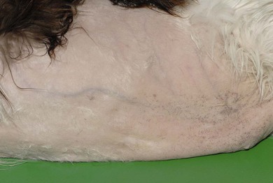
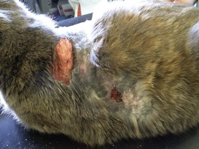
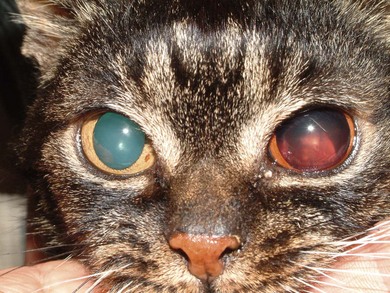
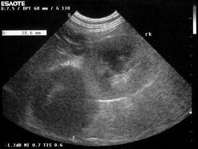
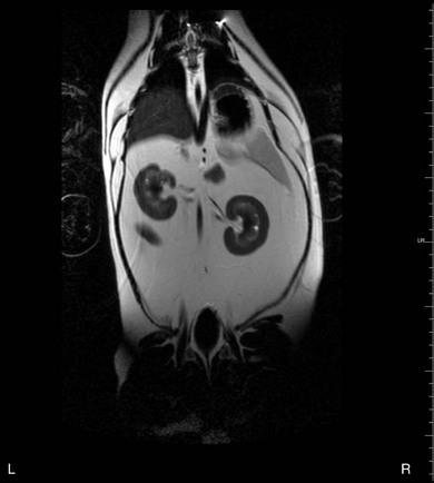
Chapter 35 The adrenal glands are found cranial to each kidney. They are cream colored as they are rich in lipids, and on ultrasound examination they have been reported to be 0.29–0.53 cm in size along their short axis.1 At surgery, the duodenal maneuver is used to identify the right side and the colonic maneuver the left. Imaging is essential both for diagnostics and patient assessment before surgery. Currently, ultrasonography is the best imaging modality for detecting adrenal masses in dogs and cats. Inflated thoracic radiographs and/or computed tomography are used to rule out metastatic disease. Abdominal ultrasound, preferably combined with CT or MRI, should be performed to look for evidence of metastatic disease within the abdominal cavity (liver, spleen, lymph nodes), but also to assess local invasion or thrombosis within the vena cava. This information can make a great difference to surgical planning and treatment advice. Invasion into the adjacent vena cava, or renal vessels, renal or liver parenchyma is possible with more aggressive neoplasms and may be a contraindication for surgery if invasion is extensive. There are few reports in the literature about surgical management of caval invasion in the cat2 and so guidelines are not available for when surgery should be undertaken, but this may be largely dictated by surgeon experience. Hyperaldosteronism may be of primary or secondary etiology. Secondary hyperaldosteronism occurs as a response to stimulation of the renin–angiotensin–aldosterone system (RAAS) and therefore may occur in any disease that stimulates the RAAS, such as dehydration, hypotension, reduced renal perfusion (most commonly secondary to renal disease), or sodium deficiency (reduced intake or increased loss). In primary hyperaldosteronism (PHA) there is an inappropriate increase in aldosterone secretion, independent of the RAAS. Unilateral adrenocortical neoplasia is the most commonly described cause of PHA in cats, with unilateral adrenal adenomas and carcinomas appearing to have approximately equal incidence. Although rare, bilateral adrenal neoplasms have also been described. Bilateral adrenal hyperplasia3 is also recognized, and in these cases medical management is advised. • Intraocular hemorrhage or acute onset blindness resulting from retinal detachment (Fig. 35-1) as a result of systemic hypertension. Figure 35-1 A cat with retinal detachment and vitreal hemorrhage, as a result of systemic hypertension associated with hyperaldosteronism. (Photo courtesy of Tim Knott, Rowe Veterinary Group, UK. Reprinted from August, JR. 2010 Consultations in Feline Internal Medicine 6e, W.B. Saunders, with permission from Elsevier.) Additional clinical signs such as polyuria, polydipsia, and polyphagia have also been reported Concurrent hyperaldosteronism and hyperprogesteronism has also been reported in some cases of adrenal neoplasia.4,5 In these cases the signs of hyperprogesteronism usually predominate, resulting in very similar clinical signs to those encountered with hypercortisolemia (Fig. 35-2). Adrenal masses are rarely visible on radiographs, but if the mass is seen it is more likely to be an adrenocortical carcinoma than an adenoma. Pulmonary metastases can occur, albeit infrequently. In all reported feline cases of primary hyperaldosteronism in which adrenal ultrasonography was performed, unilateral adrenal enlargement with evidence of an adrenal mass was identified, ranging in size from 10–46 mm in diameter (Fig. 35-3). The contralateral adrenal gland may appear normal in appearance or may be unidentifiable. It is, however, vital that the contralateral gland be assessed, because bilateral adrenal neoplasms have been reported. Ultrasonography should also be used to identify the presence and degree of invasion of the caudal vena cava by the tumor or related thrombus, and the presence of metastasis to other organs. Close association of the tumor with the caudal vena cava is usually evident. Other imaging modalities, including MRI (Fig. 35-4), CT, and saphenous venography, have been used in a small number of cases in an attempt to establish the extent of the adrenal mass before undertaking surgery. Figure 35-3 Ultrasound image demonstrating the typical hypoechoic appearance of an adrenal mass cranial to the right kidney. This was an adrenocortical carcinoma. (Reprinted from August JR. (2010) Consultations in Feline Internal Medicine, 6, W.B. Saunders, with permission from Elsevier.) Figure 35-4 T2-weighted sagittal MRI scan of the abdomen demonstrating significantly enlarged right adrenal gland. The major clinical signs are similar, whether hyperadrenocorticism results from adrenal adenoma, carcinoma or hyperplasia. Approximately 75% of cats with hyperadrenocorticism develop secondary diabetes mellitus, and the signs of uncontrolled diabetes mellitus usually predominate, namely polyuria, polydipsia, and polyphagia. Other clinical signs include lethargy, weight loss, an unkempt coat, and abdominal enlargement/pot belly appearance, although this is usually less appreciable than it is in dogs. Failure of hair regrowth, thin skin, and poor wound healing are common dermatologic features. Cushingoid cats may also exhibit severe skin fragility where the skin is so thin that it easily tears (Fig. 35-5). This is a serious complication as resultant wounds heal poorly, and it can also result in life-threatening sepsis.
Adrenal gland
Surgical anatomy
Diagnosis and general considerations
Diagnostic imaging
Surgical diseases
Hyperaldosteronism (Conn syndrome)
Etiology
Clinical features
Diagnosis
Hyperadrenocorticism (Cushing’s syndrome)
Clinical features
![]()
Stay updated, free articles. Join our Telegram channel

Full access? Get Clinical Tree


Adrenal gland