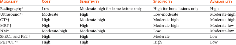
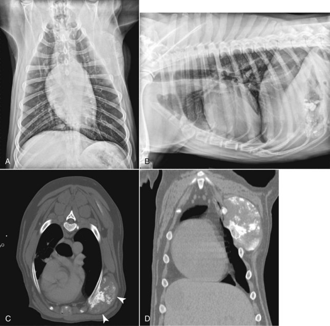
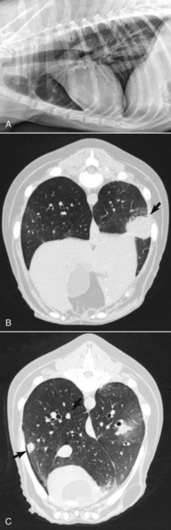
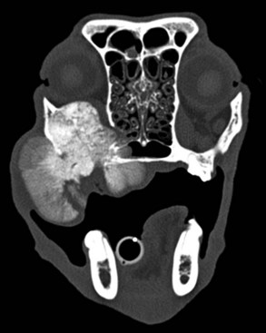
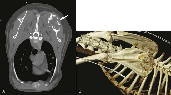
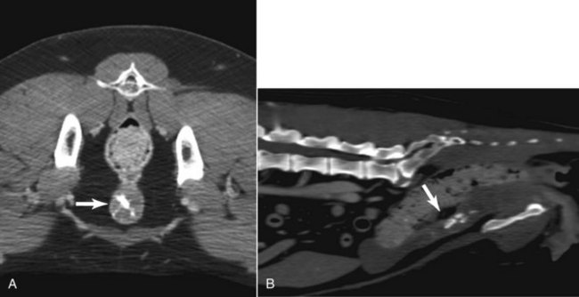
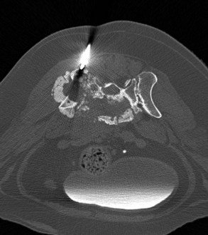
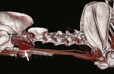
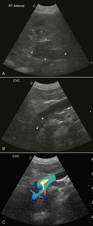
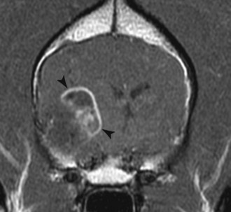
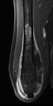
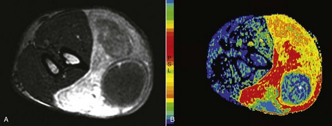
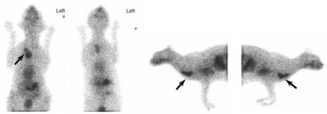
6 Diagnostic imaging plays an essential role in the management of the cancer patient. The initial diagnosis, staging, surgical and radiation treatment planning and response to therapy all involve imaging to a varying extent. Routine radiographs, ultrasound, nuclear medicine, and cross-sectional imaging in the form of computed tomography (CT) and magnetic resonance imaging (MRI) are routinely used in veterinary oncology. The choice of imaging modality depends on many factors, including the desired outcome. The biologic behavior of the tumor directs the imaging choice in cancer staging, and imaging may play an important role in guiding serial tumor biopsy during the course of therapy. The sophistication of imaging modalities continues to increase exponentially. Each modality has advantages and disadvantages with regard to cost, availability, sensitivity, specificity, and qualities of anatomic versus functional imaging (Table 6-1). Advanced molecular imaging techniques, which measure biologic processes at the cellular level,1–3 are quickly becoming commonplace in physician-based oncology and have the potential to play an important role in the tailoring of cancer therapy in veterinary patients. Conventional radiography has been the mainstay of cancer imaging for many years because of its accessibility and low cost. However, it usually is relegated to a screening test and is often followed by other imaging modalities to better differentiate and define tumor extent (Figure 6-1).4,5 Radiographic images are produced by the differential absorption of x-rays as the primary beam passes through the patient. Some x-ray photons are absorbed by the body and some pass through. Absorption depends on the thickness, physical density, and effective atomic number of the tissues of the patient’s body. The x-rays that are not absorbed reach the radiographic film and determine the blackness and gray scale of the image. Two of the strengths of radiography are the global information it provides and its excellent utility for bone imaging, especially the appendicular skeleton.6 Thoracic and abdominal radiographs are excellent screening tools for feline and canine lymphoma patients to determine thoracic lymph node and pulmonary involvement, as well as liver, spleen, and abdominal lymph node neoplastic spread.7,8 Radiography’s greatest weaknesses are the superimposition of overlying structures and that only a few radiographic opacities are depicted. CT and MRI have replaced radiography for imaging of the head and, in some circumstances, the axial skeleton.9–12 As does radiography, CT relies on the physical density differences between tissues to form the image. Unlike radiography, CT portrays slices of the patient without superimposition of structures because the images are computer generated and the gray scale display is superior. Although thoracic radiographs are routinely used as a screening method for evaluation of primary and metastatic tumors of the lung, mediastinum, and ribs, CT provides superior information for characterizing and anatomically localizing thoracic lesions for their diagnosis and treatment (see Figure 6-1). Compared with radiography, CT is more sensitive for identifying pulmonary nodules (Figure 6-2), mediastinal lymphadenopathy, and pleural and other masses.5,13–17 CT should be used to ascertain the full extent of pulmonary nodules from metastatic disease and when a primary lung tumor has been identified to evaluate for intrathoracic metastases and tracheobronchial lymphadenopathy. CT is more sensitive than radiography to osteolysis and osteoproduction associated with neoplasia, and its three-dimensional (3D) information is especially useful for skeletal structures such as the sinonasal region, orbit (Figure 6-3), ear canals, and skeleton (Figure 6-4).9 CT is also useful to determine the origin and extent of abdominal mass lesions, and compared to ultrasonography, CT can better document the relationship of a mass with surrounding anatomic structures (Figure 6-5).18,19 Infiltrative muscular lesions such as infiltrative lipomas and soft tissue sarcomas are routinely imaged with CT for both surgical and radiotherapy planning.12,20 A contrast-enhanced scan is essential during CT to improve visualization of tumor margins, especially for infiltrative tumors such as feline vaccine–associated sarcomas.21 CT is exceedingly useful for both surgical and radiotherapy planning. Although MRI has better tissue differentiation, CT is used most often for radiotherapy treatment planning because there is no image distortion and the physical tissue density is available for input into treatment planning computers.22,23 It is extremely important to position the radiotherapy patient for the CT scan done for radiation treatment planning in a manner that can reliably be repeated during therapy. This will ensure the treatment is delivered as planned. CT is also amenable to obtaining image-guided biopsy of masses that are not readily obtained with ultrasound guidance, and is particularly helpful for thoracic, brain, spinal, and skeletal lesion biopsy (Figure 6-6).24–28 Contrast-enhanced CT angiography (CTA) is now also becoming more routine due to the increasing availability of multidetector CT scanners in veterinary medicine. CTA allows detection of tumor vascular invasion and can also depict tumor vascular supply for interventional therapies (Figure 6-7). Dynamic multiphase CT can also improve detection of metastases and small tumors such as insulinomas and may distinguish benign from malignant hypervascular primary hepatic tumors.29–32 Tumor perfusion, vascular permeability, and tumor blood volume can predict tumor aggressiveness through the use of dynamic contrast-enhanced CT, which estimates tumor wash-in and wash-out contrast kinetics.33,34 Ultrasound is superior to radiographs in instances of pleural or peritoneal effusion due to the loss of visceral detail that occurs on radiographs as the fluid will efface the margins of organs.35 Ultrasound is also useful in guidance for biopsy or fine-needle aspiration of an observed abnormality. It is more sensitive than survey radiographs for detecting abdominal lymphadenopathy36–38 and size, shape, margins, heterogeneity, perinodal fat appearance, and vascular pattern of lymph nodes are useful in determining malignancy.38–42 However, radiographs are superior for detection of bony invasion that may be associated with malignant medial iliac lymph node enlargement, often seen with tumors of the urinary bladder, prostate,43 and anal glands. Ultrasound is sensitive in the detection of adrenal abnormalities, including vessel invasion, which is usually associated with malignancy (Figure 6-8).19,44 Ultrasound is routinely used to stage and monitor cats and dogs with mast cell disease, which has a varied appearance.45,46 It is used extensively in patients with gastrointestinal disease for initial diagnosis and monitoring of therapy.47–52 Ultrasonography is sensitive for lesion detection, but it is not specific for disease etiology. Many studies have attempted unsuccessfully to differentiate benign from malignant lesions based on sonographic appearance.44,46,50,53–55 Therefore biopsy or fine-needle aspirates of lesions are necessary. Ultrasound-guided sampling of tissue can be performed quickly, accurately, and safely.56–60 A caveat is the potential seeding of tumor cells from ultrasound-guided percutaneous sampling of transitional cell carcinoma.61 Advances in ultrasound equipment and the development of sonographic contrast agents are increasing the specificity of ultrasound.62 Doppler techniques are used to assess tumor vasculature, which is found generally to be tortuous with high velocity as compared to normal tissues.40,42,63,64 Tissue harmonic imaging transmits at one frequency and receives at twice that frequency. This technique has advantages over fundamental imaging.62,65 Harmonic imaging and/or ultrasound contrast agents have been used to differentiate benign from malignant lesions64–76 and continued exploration of this imaging modality is warranted. MRI is an advanced imaging technique that provides superb soft tissue images and sensitive detection of pathology based on properties of hydrogen atoms when placed in magnetic and radiofrequency fields. Images can be acquired directly in any plane with MRI, compared to CT in which sagittal and dorsal images are made by reconstructing transverse image data. MRI is the imaging modality of choice for the evaluation of the central nervous system, and the characteristics of the common canine and feline neurologic tumors are now well described (Figure 6-9).77–82 MRI provides better anatomic detail and more sensitive detection of neuropathology than CT but is less useful for assessing cortical bone because of the number and characteristics of hydrogen protons in bone. Bone landmarks, areas of mineralization, and periosteal bone production are not as obvious with MRI as with CT. Nevertheless, MRI provides excellent anatomic detail and sensitive detection of infiltrative diseases affecting the musculoskeletal soft tissues, including joints, ligaments and tendons, and bone marrow, and is most accurate in determining the extent of canine appendicular osteosarcoma for limb-sparing procedures (Figure 6-10).83 As advanced MR equipment becomes more common in veterinary medicine, MRI’s use in cancer diagnosis and staging will increase because of its value in determining tumor morphology, margins, characteristics, and composition.32,84 MRI is excellent for tumors of the head, neck, and torso, although special measures are sometimes needed to minimize respiratory motion in certain anatomic sites.85,86 Whole-body MR is under evaluation as a sensitive method for cancer staging that is superior to CT and scintigraphy, and in human clinical trials it can rival the results of positron-emission tomography-CT (PET-CT) without the use of ionizing radiation.87 Whole-body MRI does require ultrafast imaging methods, so it would be limited to veterinary sites having newer and high-field MR instruments.84,87 Although CT scans are most often used for radiation therapy planning, MRI provides advantages in select situations, including the ability to anatomically define small at-risk structures such as the optic chiasm and to contour margins of lesions where beam-hardening artifact is problematic with CT.84 MRI is also quite useful for detecting residual or recurring tumors due to its sensitivity to early disease. New MRI techniques are being developed that allow the study of tumor physiology and metabolism and valuable information about treatment response. Dynamic contrast-enhanced MRI provides information about tumor vascularity, perfusion, and angiogenesis that has been shown to have predictive value for treatment response and outcome (Figure 6-11).88,89 Diffusion MRI evaluates the mobility of tumor water molecules as a function of cell density and tissue architecture, with an increase in tumor diffusion indicating a positive treatment response and cell death.32,88,90,91 In vivo MR spectroscopy for measuring malignant metabolic biomarkers such as choline relative to normal surrounding tissue is moving from the research realm into human clinical use for brain and prostatic cancer and also has future potential for veterinary diagnosis.84,92 Diagnostic nuclear medicine, or scintigraphy, involves the administration of radiopharmaceuticals that localize to an area of interest in the body by physiologic processes. Images obtained from nuclear medicine studies do not provide the anatomic detail attainable with other imaging techniques; however, the functional dependence on physiologic processes adds important information. Technetium-99m (99mTc) is the most commonly used radionuclide because it has excellent imaging qualities and a short half-life (6 hours) and is easily bound to localizing pharmaceuticals. Bone scintigraphy using 99mTc methylene diphosphonate (99mTc-MDP) is frequently used in veterinary medicine because it is a simple, sensitive, and noninvasive method of evaluating the entire skeleton.93 Other commonly used nuclear imaging studies include renal, thyroid, lung, and liver scintigraphy (Figure 6-12). Parathyroid scintigraphy has been performed using dual-phase imaging94 but due to lack of sensitivity was not recommended for identification of abnormal parathyroid glands in hypercalcemic dogs. At this author’s (LJF) institution, ultrasound is used primarily to locate parathyroid masses in hypercalcemic dogs and cats.95,96
Imaging in Oncology
Imaging Modalities
Computed Tomography
Ultrasonography
Magnetic Resonance Imaging
Nuclear Medicine
![]()
Stay updated, free articles. Join our Telegram channel

Full access? Get Clinical Tree


Imaging in Oncology